This is a student handbook—a concise, high-yield reference designed to help medical students understand the difficult principles of hematology. It brings together two companion guides: benign (non-malignant) hematology and malignant hematology. It is meant to sit beside your lectures and textbooks as a study aid, not to replace them.
How the handbook is organised
The handbook is divided into two books. Book One — Benign Hematology covers the foundations (how blood is made and measured), the anemias, the non-malignant white cell disorders, bleeding disorders, thrombosis and anticoagulation, and transfusion medicine. Book Two — Malignant Hematology covers the blood cancers: the founding concepts, the myeloid and lymphoid malignancies, and the cross-cutting treatments of transplantation, supportive care, immunotherapy, and cellular therapy. Chapters are numbered within each book. Every chapter follows the same structure — the big idea, the substance, pitfalls and pearls, a case, and a quick review — so the framework always comes first and the diseases follow as applications of it.
A note on authorship and status
This handbook was created with the assistance of artificial intelligence (Claude, made by Anthropic) as a free study aid for students. It is NOT a peer-reviewed publication, a textbook, or an official clinical reference, and it should not be cited as one. It has not undergone formal editorial or expert review. The content should always be cross-checked against standard textbooks, current clinical guidelines, and your own faculty before being relied upon for examinations or, especially, for patient care. Drug doses and thresholds are deliberately kept general, medicine changes over time, and errors are possible in any AI-assisted material. Where this handbook and an authoritative source disagree, trust the authoritative source.
PLEASE NOTE
A student-made reference handbook, created with AI assistance (Claude).
Not a publication, not peer-reviewed, and not for citation.
A study aid only — always verify against textbooks, guidelines, and faculty before any clinical use.
Section One · Benign Hematology
A High-Yield Guide for Medical Students
PART I · FOUNDATIONS
How to think about blood before you memorize the diseases
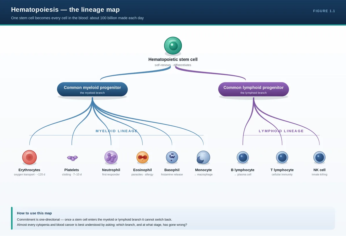
All blood cells descend from a single, rare, self-renewing hematopoietic stem cell (HSC) in the bone marrow. As that stem cell divides, its offspring progressively commit down one of two great lineages—myeloid or lymphoid—losing the ability to become anything else. Almost every hematologic disease is a disturbance at a specific point on this lineage map, so knowing the map tells you where a disease lives.
Key pathophysiology
Blood has two parts. The formed elements (red cells, white cells, platelets) make up about 45% of volume; plasma, the protein-rich fluid phase carrying clotting factors and immunoglobulins, makes up the rest. Spun in a tube, red cells sink, plasma rises, and a thin grey buffy coat of white cells and platelets sits between them.
Hematopoiesis is the continuous manufacture of these cells—roughly 100 billion new cells per day. It is sustained by HSCs, which have two defining properties: self-renewal (making more stem cells, so the supply never runs out) and multipotency (the ability to generate every blood lineage). HSCs sit quietly in specialized marrow niches; their progeny amplify enormously as they mature.
Commitment is one-directional. The HSC gives rise to a common myeloid progenitor and a common lymphoid progenitor. The myeloid branch produces red cells, platelets (from megakaryocytes), neutrophils, eosinophils, basophils, and monocytes. The lymphoid branch produces B cells, T cells, and natural killer cells. Lineage is steered by growth factors (e.g., erythropoietin for red cells, thrombopoietin for platelets, G-CSF for neutrophils) and master transcription factors.
The lineage map at a glance
Progenitor branch
Mature cells produced
Primary job
Erythroid (myeloid)
Red blood cells
Oxygen transport
Megakaryocytic (myeloid)
Platelets
Primary hemostasis
Granulocyte/monocyte (myeloid)
Neutrophils, eosinophils, basophils, monocytes
Innate immunity
Lymphoid
B cells, T cells, NK cells
Adaptive immunity
Clinical clues
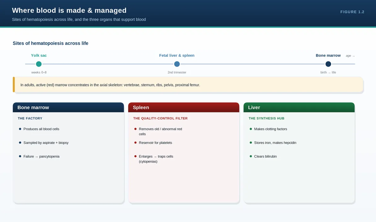
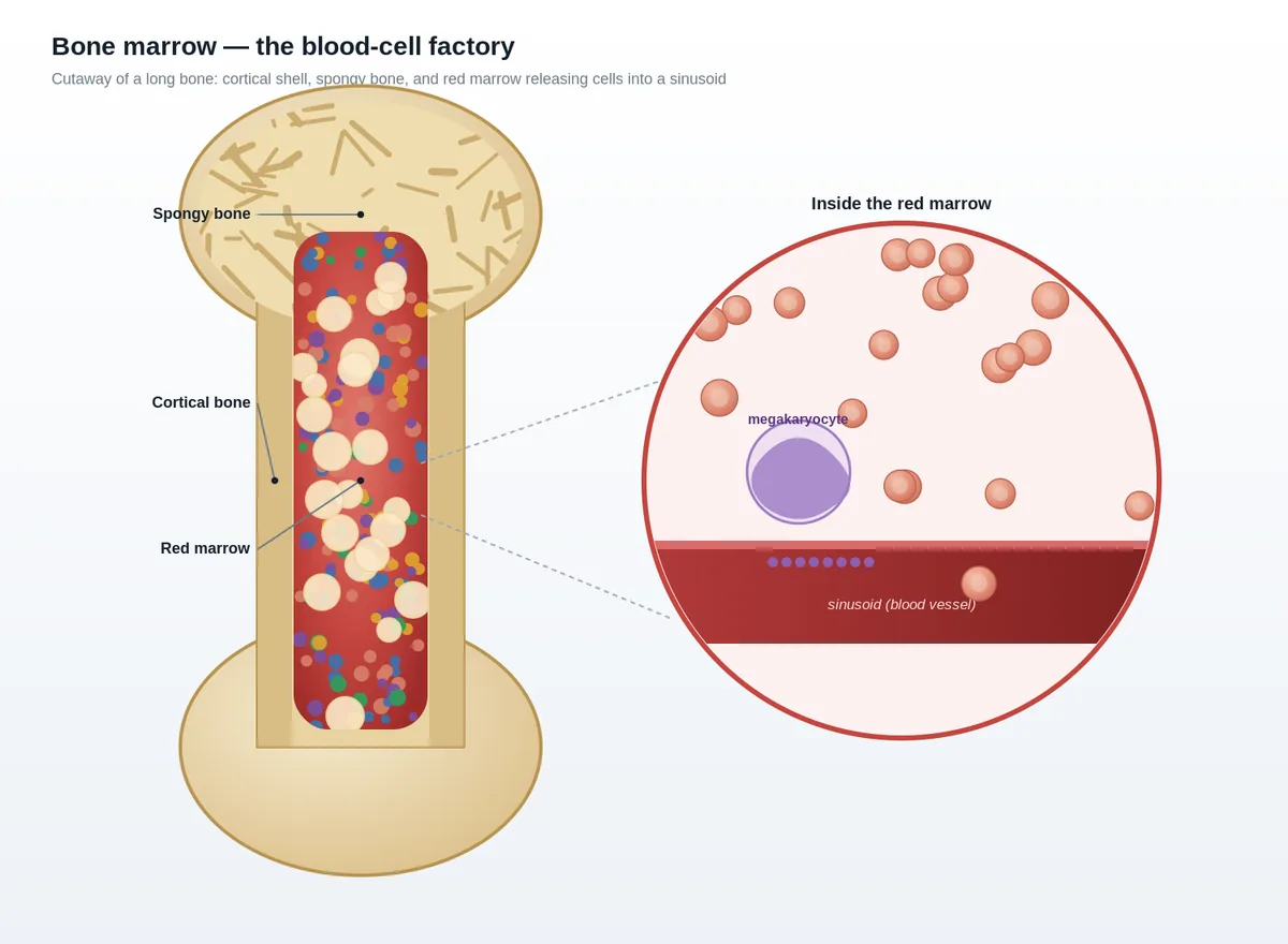
Where blood is made shifts across life: the yolk sac in early embryo, then the fetal liver and spleen, and from the third trimester onward the bone marrow. In adults, active (red) marrow is confined to the axial skeleton—vertebrae, sternum, ribs, pelvis, and proximal femora/humeri. This is why a bone marrow biopsy is taken from the posterior iliac crest, and why marrow stress can cause extramedullary hematopoiesis in the liver and spleen, producing hepatosplenomegaly.
Diagnostic approach
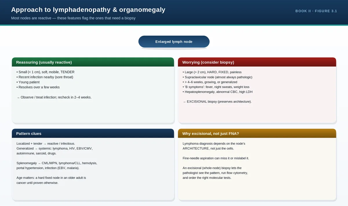
Three tissues support blood and recur throughout the guide. The bone marrow is the factory. The spleen is the quality-control filter that removes aged or abnormal red cells and is a reservoir for platelets—remove it and platelet counts rise and abnormal red cell forms (Howell-Jolly bodies) appear. Lymph nodes and thymus are where lymphocytes mature and mount immune responses, which is why lymphoid malignancies present as lymphadenopathy.
PITFALLS & PEARLS
“Stem cell” means self-renewal PLUS multipotency. A progenitor that has lost self-renewal is not a stem cell, even if it still makes several lineages.
Red marrow retreats to the axial skeleton with age—chronic anemia can re-expand marrow into long bones and even the skull (e.g., thalassemia).
The spleen both removes and stores: post-splenectomy you see thrombocytosis and Howell-Jolly bodies on the film.
Erythropoietin is made by the kidney—a key reason chronic kidney disease causes anemia.
CASE VIGNETTE
A 24-year-old with lifelong severe anemia has prominent cheekbones, a skull X-ray showing a “hair-on-end” appearance, and marked splenomegaly.
Concept: chronic marrow drive has expanded hematopoiesis into the facial bones and skull, and extramedullary hematopoiesis has enlarged the spleen—exactly what the lineage-and-location framework predicts in severe thalassemia.
QUICK REVIEW
Blood = formed elements (RBC, WBC, platelets) + plasma.
One HSC → myeloid and lymphoid branches → all blood cells.
HSC defining traits: self-renewal + multipotency.
Adult red marrow = axial skeleton; biopsy from posterior iliac crest.
Spleen filters and stores; EPO from kidney, TPO drives platelets, G-CSF drives neutrophils.
Hematopoiesis — the lineage mapWhere blood is made & managedBone marrow — anatomical cutaway
2Red Cell, White Cell, and Platelet Physiologytop ↑
THE BIG IDEA
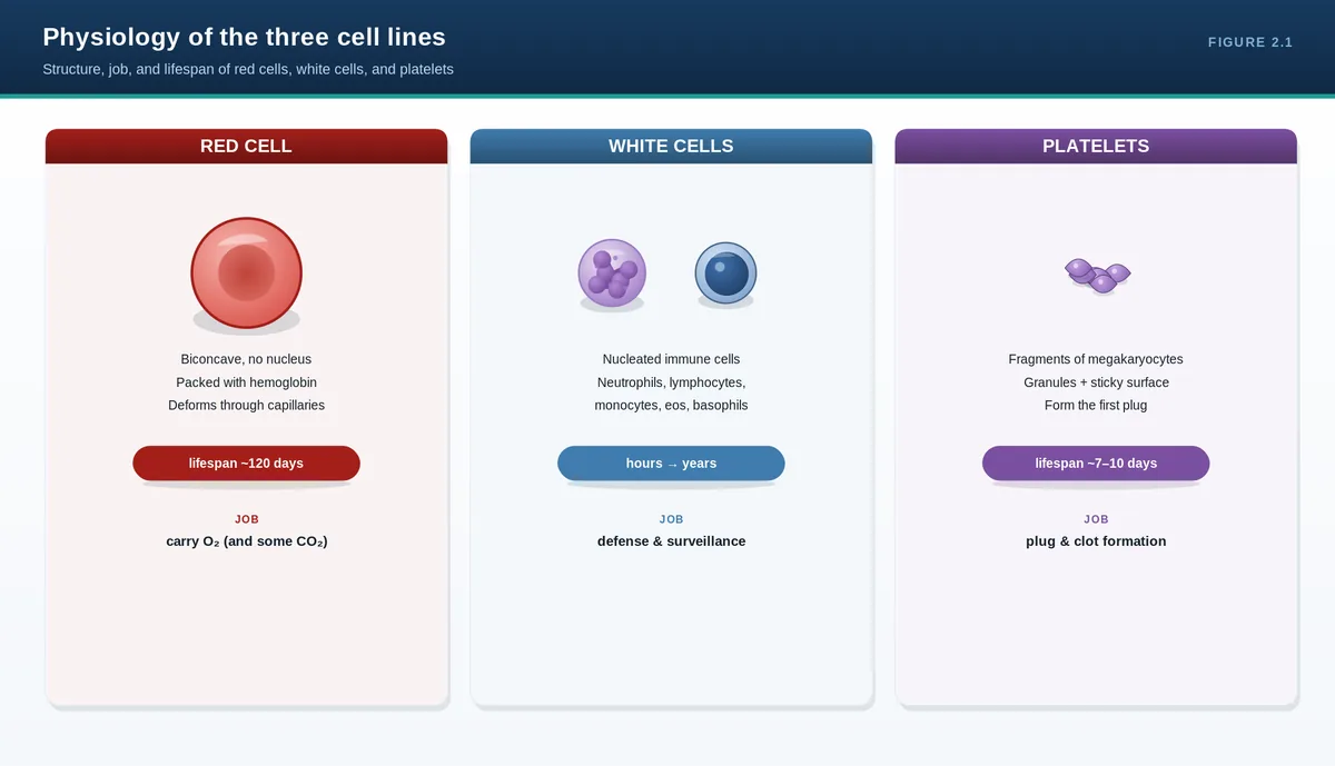
Each cell’s structure is built for one job. Red cells are biconcave, hemoglobin-filled sacs with no nucleus—optimized to carry oxygen and squeeze through capillaries for ~120 days. White cells are mobile defenders, each subtype tuned to a different threat. Platelets are anucleate cell fragments that plug vascular breaches in seconds. Understanding the normal job makes every disease a recognizable deviation from it.
Red cells and hemoglobin
A red cell is essentially a deformable bag of hemoglobin. Its biconcave disc shape maximizes surface area for gas exchange and lets it bend through capillaries narrower than itself. It has no nucleus or mitochondria, so it cannot repair itself and relies on glycolysis for energy—features that matter when we discuss membrane and enzyme disorders later.
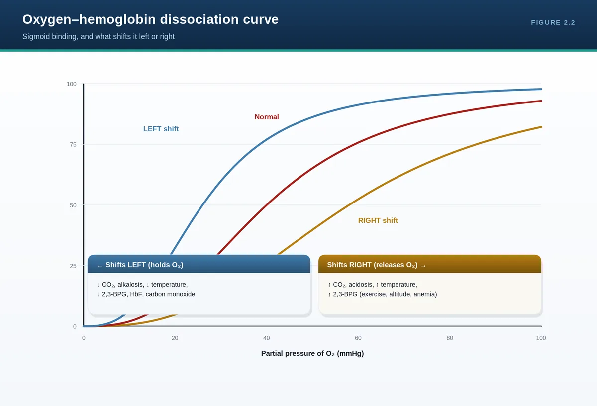
Hemoglobin is four globin chains, each cradling a heme group with one iron atom that binds one oxygen molecule. Normal adult hemoglobin is HbA (two alpha, two beta chains). Oxygen binding is cooperative, producing the sigmoid oxygen-dissociation curve: hemoglobin loads oxygen avidly in the lungs and releases it in tissues. The curve shifts right (releasing more oxygen) with acidosis, raised CO2, fever, and raised 2,3-BPG—the body’s way of delivering more oxygen where metabolism is high.
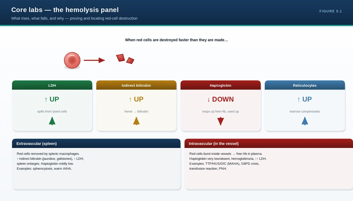
Red cells live about 120 days. Aged cells are removed by splenic macrophages, which recycle iron and break heme down to bilirubin. This is why hemolysis (premature destruction) raises unconjugated bilirubin and LDH while consuming haptoglobin—the lab signature you will use repeatedly.
White cells
Cell
Approx. share of WBCs
Main role
Neutrophil
40–60%
First responder to bacterial infection; phagocytosis
Histamine release; allergic and inflammatory reactions
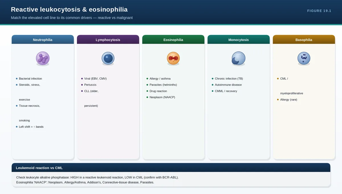
Neutrophils dominate and are short-lived (hours in blood), so their numbers swing fast with infection or steroids. Lymphocytes are long-lived and carry immunologic memory. Knowing which cell rises tells you which process is at work: neutrophilia suggests bacterial infection or inflammation, lymphocytosis suggests viral infection, and eosinophilia points to allergy, parasites, or drug reactions.
Platelets and primary hemostasis
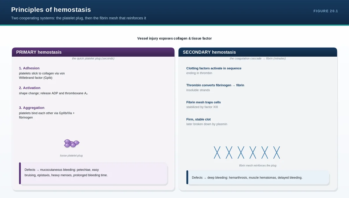
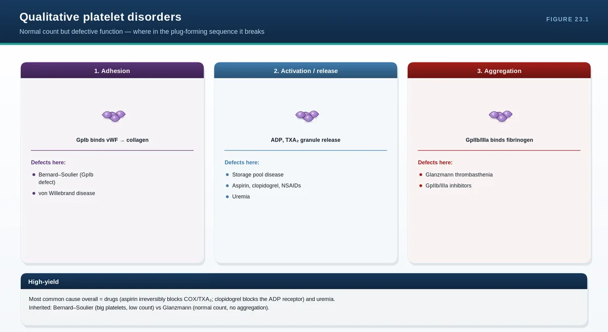
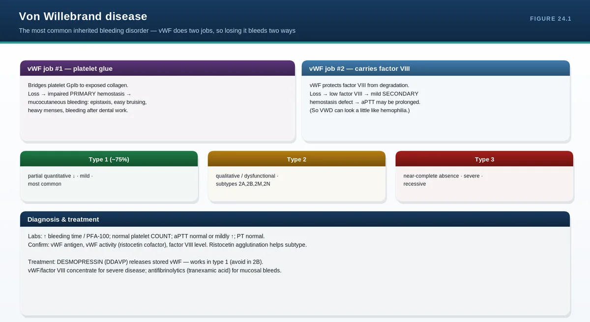
Platelets are fragments shed from marrow megakaryocytes under the control of thrombopoietin (made by the liver). They circulate for 7–10 days. When a vessel is injured, platelets adhere to exposed collagen—using von Willebrand factor as molecular glue—then activate, change shape, release granules, and aggregate into a plug. This is primary hemostasis. Defects here (low platelet count or poor platelet function, including von Willebrand disease) cause mucocutaneous bleeding: petechiae, bruising, epistaxis, and heavy periods.
PITFALLS & PEARLS
No nucleus, no mitochondria: the mature red cell cannot repair damage—hence its finite ~120-day lifespan and vulnerability in enzyme/membrane disorders.
Right shift of the O2 curve = better tissue unloading (acid, CO2, heat, 2,3-BPG). Remember “exercising muscle wants oxygen.”
Hemolysis lab triad: ↑ unconjugated bilirubin, ↑ LDH, ↓ haptoglobin (plus reticulocytosis).
Mucocutaneous bleeding ⇒ think primary hemostasis (platelets/VWF). Deep joint/muscle bleeds ⇒ think secondary hemostasis (coagulation factors).
CASE VIGNETTE
A 30-year-old woman reports easy bruising, frequent nosebleeds, and heavy menstrual periods. Joint bleeds are absent.
Concept: the pattern is mucocutaneous, pointing to a primary-hemostasis problem (platelet number/function or von Willebrand factor)—not a coagulation-factor deficiency, which would more often cause deep bleeding.
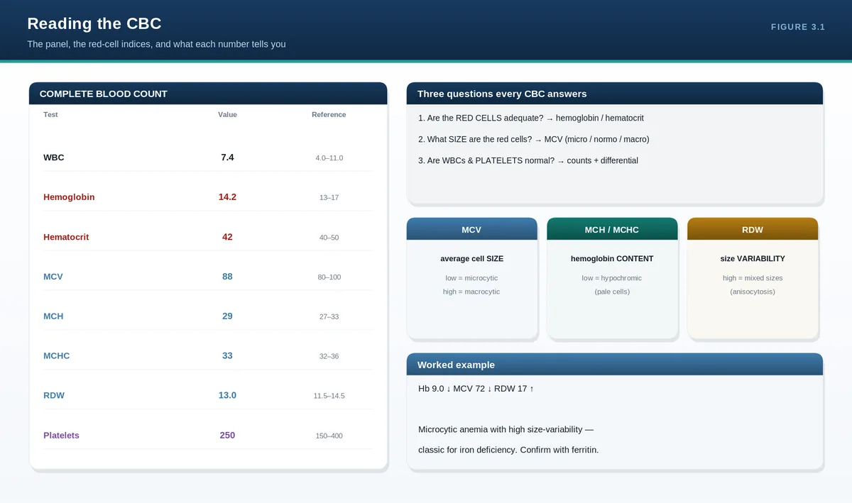
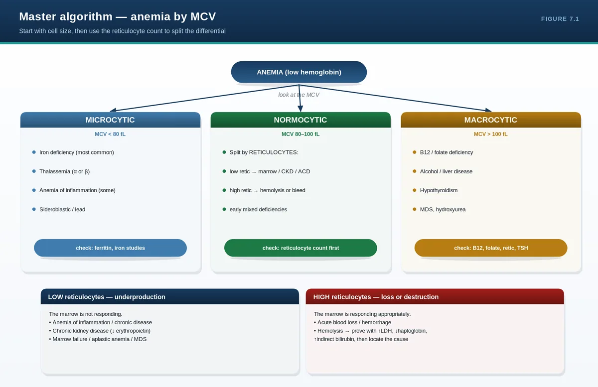
The CBC is three production lines reported on one page: red cells, white cells, and platelets. Don’t read it as a list of numbers—read it as three questions. Is each line too high, too low, or normal? And for any abnormal line, are the cells the right size and is the marrow responding? Two numbers—the MCV and the reticulocyte count—turn anemia from a long differential into a short one.
Total white cells; interpret with the differential
Platelets
150–400 ×10⁹/L
Primary hemostasis capacity
Diagnostic approach: the two-number method for anemia
Step 1 — size (MCV). Microcytic (<80 fL) anemias are dominated by iron deficiency and thalassemia (and, less often, anemia of chronic disease and sideroblastic anemia). Normocytic (80–100 fL) anemias include acute blood loss, hemolysis, anemia of chronic disease, and marrow problems. Macrocytic (>100 fL) anemias split into megaloblastic (B12/folate deficiency) and non-megaloblastic (alcohol, liver disease, hypothyroidism, myelodysplasia, reticulocytosis).
Step 2 — response (reticulocytes). A high reticulocyte count means the marrow is working hard to replace lost cells—pointing to bleeding or hemolysis. A low or inappropriately normal reticulocyte count means the marrow is failing to respond—pointing to a production problem (nutrient deficiency, marrow disease, or lack of erythropoietin). The reticulocyte count is what separates “losing/destroying cells” from “not making enough.”
The RDW adds nuance: it is high when a population of cells varies in size. A high RDW with microcytosis favors iron deficiency (a mix of old normal and new small cells), whereas thalassemia trait classically shows a low/normal RDW with uniformly small cells and a near-normal red cell count.
Clinical clues: common whole-CBC patterns
Microcytic anemia, high RDW: iron deficiency until proven otherwise—find the bleeding source.
Pancytopenia (all three lines low): marrow failure, infiltration, megaloblastic anemia, or hypersplenism.
High WBC with left shift, normal Hb/platelets: reactive neutrophilia (infection, inflammation, steroids).
High Hb, WBC, and platelets together: suspect a myeloproliferative process
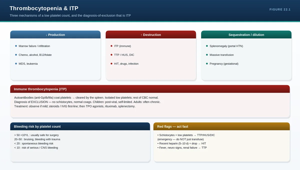
Isolated thrombocytopenia in a well patient: think ITP, but always confirm it is not a lab artifact (clumping).
PITFALLS & PEARLS
Always confirm a surprising low platelet count with a film: EDTA-induced platelet clumping causes spurious (pseudo-)thrombocytopenia.
A normal MCV does not exclude mixed deficiency—iron + B12 deficiency can produce a normal “average” size with a high RDW.
Reticulocytes are reported as a percentage; in severe anemia correct for the low red cell mass (reticulocyte index) so you don’t overcall a good marrow response.
Hematocrit ≈ 3 × hemoglobin (g/dL). A big mismatch suggests a lab or sampling problem.
CASE VIGNETTE
A 45-year-old woman: Hb 9.0 g/dL, MCV 72 fL, RDW 18%, platelets 450 ×10⁹/L, reticulocytes low.
Reasoning: microcytic + high RDW + low reticulocytes + reactive thrombocytosis = iron deficiency. The next step is iron studies and a search for blood loss (menstrual or gastrointestinal).
QUICK REVIEW
Read the CBC as three lines; for any abnormal line ask: right number? right size? marrow responding?
MCV classifies anemia: micro (<80), normo (80–100), macro (>100).
Reticulocytes: high = bleeding/hemolysis; low = production problem.
High RDW + microcytosis → iron deficiency; low/normal RDW + microcytosis → thalassemia trait.
Confirm unexpected low platelets with a film (rule out clumping).
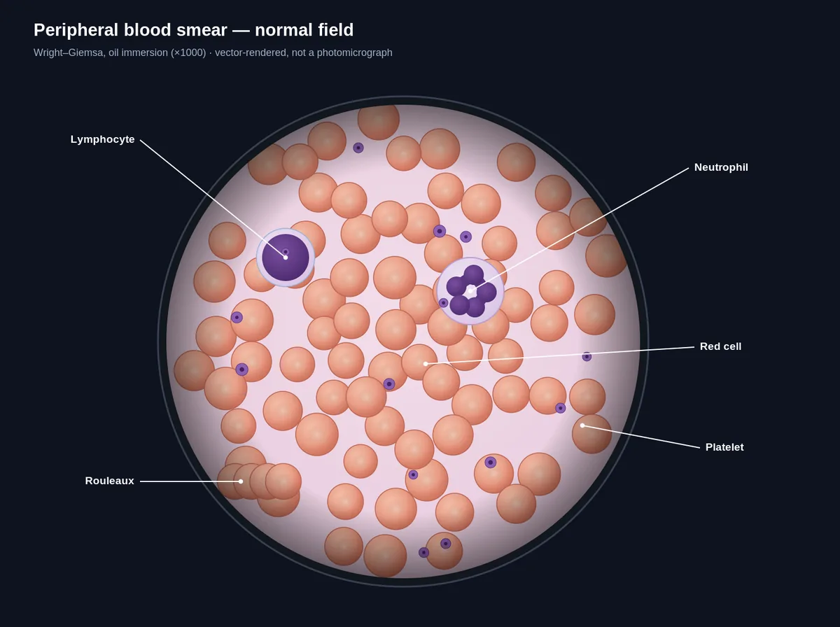
The CBC counts cells; the film lets you look at them. Numbers tell you something is wrong—the film often tells you what. A handful of morphologies are so specific that spotting one essentially makes the diagnosis. Learn to describe a film systematically (red cells, white cells, platelets) and you will never freeze when an examiner pushes one in front of you.
When to look at the film
Request a film whenever the CBC is unexpectedly abnormal, when hemolysis is suspected, when there is unexplained cytopenia or leukocytosis, or when the analyzer flags abnormal cells. The film is essential—not optional—in any suspected hemolytic anemia, leukemia, or microangiopathy.
A structured way to read red cells
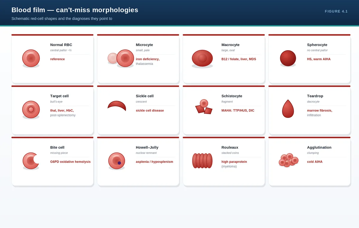
Describe four things in order: size, shape, color, and inclusions. Size gives microcytic, normocytic, or macrocytic; mixed sizes are anisocytosis. Color refers to central pallor—pale (hypochromic) cells suggest low hemoglobin content as in iron deficiency. Shape and inclusions are where the high-yield clues live.
Can’t-miss red cell morphologies
Finding
What it looks like
Think of…
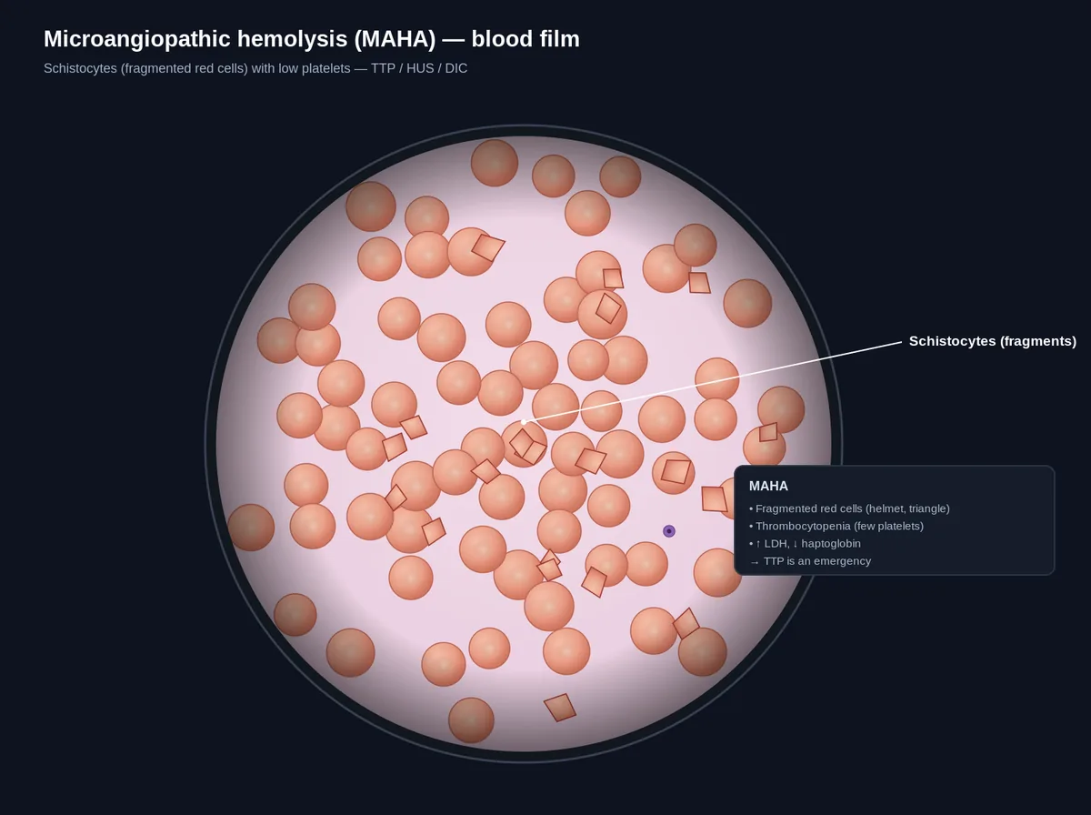
Schistocytes
Fragmented, helmet-shaped cells
Microangiopathic hemolysis (TTP, HUS, DIC)
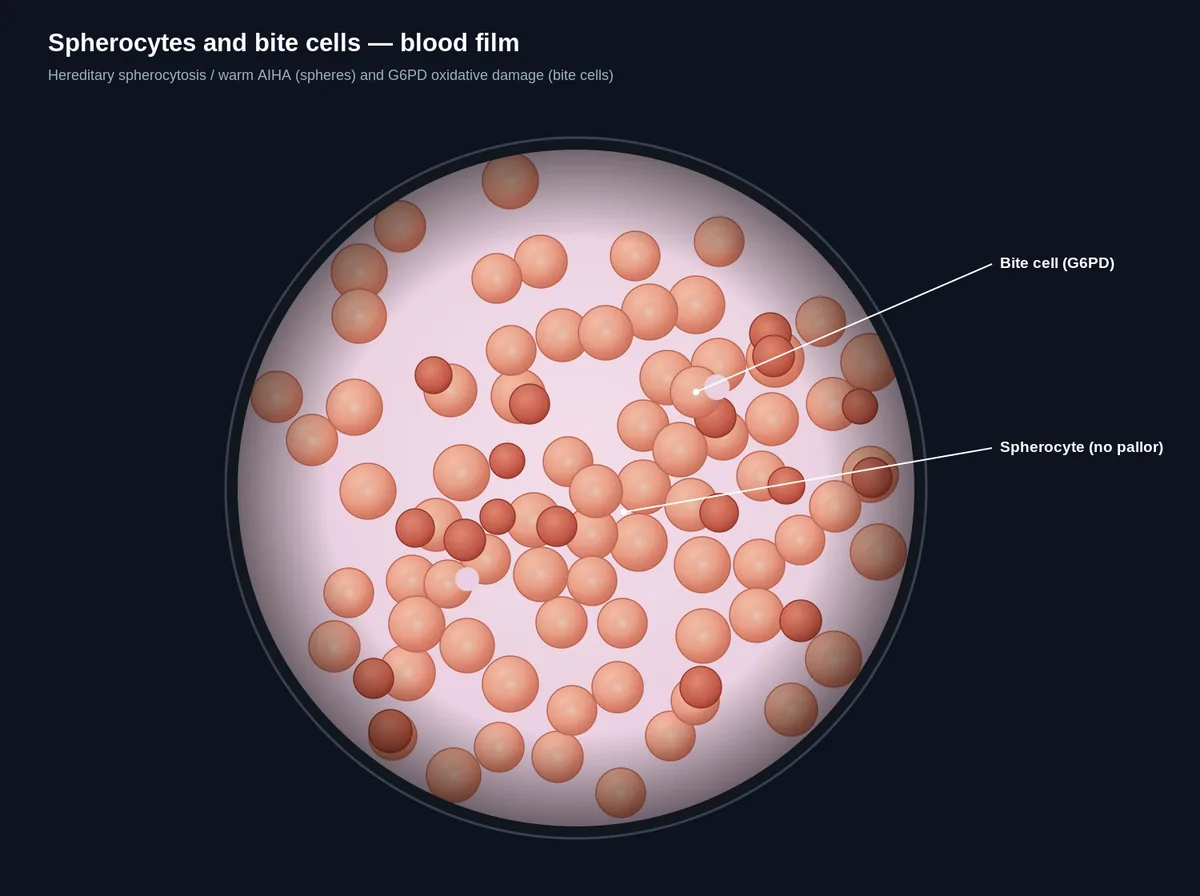
Spherocytes
Round, dense, no central pallor
Hereditary spherocytosis; autoimmune hemolysis
Target cells
Bullseye of central staining
Thalassemia, liver disease, post-splenectomy
Sickle cells
Crescent/sickle-shaped
Sickle cell disease
Tear-drop cells
Tear-shaped (dacrocytes)
Marrow fibrosis/infiltration
Howell-Jolly bodies
Single dark nuclear remnant
Hyposplenism/post-splenectomy
Blasts
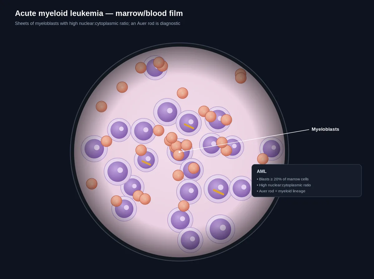
Large immature cells; Auer rods
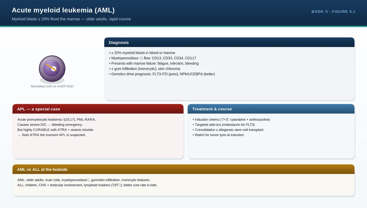
Acute leukemia
Hypersegmented neutrophils
Neutrophil with ≥6 lobes
B12/folate (megaloblastic) deficiency
White cells and platelets on the film
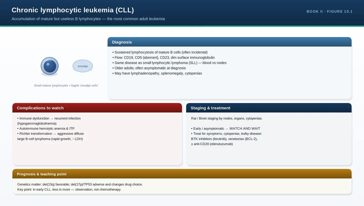
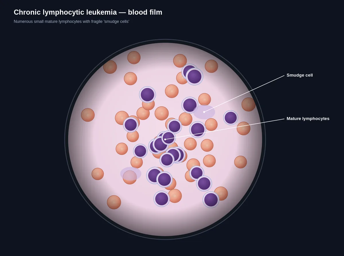
Confirm the differential and look for immaturity. A “left shift” (band forms, metamyelocytes) accompanies infection or marrow stress. Blasts or Auer rods mean acute leukemia until proven otherwise. Smudge cells suggest chronic lymphocytic leukemia. For platelets, the film confirms true numbers (clumping causes falsely low counts) and shows large or giant platelets, which suggest rapid turnover or certain inherited disorders.
PITFALLS & PEARLS
Schistocytes + thrombocytopenia is a red flag for a thrombotic microangiopathy—an emergency (think TTP).
Spherocytes have two main causes: hereditary spherocytosis and warm autoimmune hemolysis—use the direct antiglobulin (Coombs) test to separate them.
Howell-Jolly bodies on a film mean the spleen isn’t doing its job—ask why (splenectomy, sickle autosplenectomy, celiac).
Hypersegmented neutrophils can appear before the MCV rises—an early clue to megaloblastic anemia.
CASE VIGNETTE
A 28-year-old woman presents with confusion, fever, bruising, Hb 8 g/dL, platelets 18 ×10⁹/L. The film shows numerous schistocytes.
Reasoning: microangiopathic hemolysis plus severe thrombocytopenia is thrombotic thrombocytopenic purpura until proven otherwise—a medical emergency requiring urgent plasma exchange. The film made the diagnosis.
QUICK REVIEW
Film answers “what” when the CBC says “something’s wrong.”
Describe red cells by size, shape, color, inclusions.
Schistocytes → microangiopathy; spherocytes → HS or AIHA; targets → thalassemia/liver; sickle cells → SCD.
Howell-Jolly bodies → hyposplenism; hypersegmented neutrophils → megaloblastic; blasts/Auer rods → acute leukemia.
Always use the film to confirm unexpected platelet counts.
Blood film — can't-miss morphologiesPeripheral blood smear — normal field
Hematology runs on a few panels, each answering one question. Iron studies ask, “is there enough iron and where is it?” The hemolysis panel asks, “are red cells being destroyed?” Hemoglobin electrophoresis asks, “which hemoglobins are present?” The coagulation screen asks, “which arm of clotting is broken?” Learn the question each panel answers and interpretation becomes logical, not memorized.
Iron studies
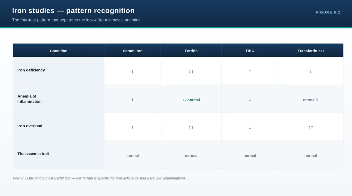
Four results work together. Ferritin reflects body iron stores—low ferritin is the single most specific test for iron deficiency. Serum iron is the iron in transit. Transferrin (or total iron-binding capacity, TIBC) is the transport protein, which rises in deficiency as the body tries to capture more iron. Transferrin saturation (iron ÷ TIBC) falls in deficiency. The trap: ferritin is an acute-phase reactant and rises with inflammation, infection, and liver disease, so a “normal” ferritin can hide iron deficiency in a sick patient.
Ferritin
Serum iron
TIBC
(=transferrin)
Transferrin sat. (Iron/TIBC)
Iron deficiency
Low
Low
High
Low
Anemia of chronic disease
Normal/High
Low
Low
Low/Normal
Iron overload
High
High
Low/Normal
High
The hemolysis panel
To prove hemolysis, look for evidence of red cell breakdown plus a marrow response. Breakdown raises LDH (released from lysed cells) and unconjugated bilirubin (heme catabolism) and consumes haptoglobin (which mops up free hemoglobin, so it falls). The marrow responds with reticulocytosis. Once hemolysis is confirmed, the direct antiglobulin (Coombs) test asks whether it is immune-mediated: positive points to autoimmune hemolysis; negative points to non-immune causes (membrane, enzyme, or mechanical).
Intravascular hemolysis (destruction within vessels): very low haptoglobin, hemoglobinuria, markedly raised LDH—seen in microangiopathy, transfusion reactions, severe G6PD crises.
Extravascular hemolysis (destruction in spleen/liver): raised bilirubin, splenomegaly, spherocytes—seen in hereditary spherocytosis and warm autoimmune hemolysis.
Hemoglobin electrophoresis
Electrophoresis and HPLC separate hemoglobins by type and quantify them, confirming hemoglobinopathies and thalassemias. Because this test is so central to red cell disorders, it has its own chapter that follows (Chapter 6). For now it is enough to know it is the confirmatory step after a suggestive CBC and film—microcytosis with normal iron, or sickle cells on the film—and that it must always be read together with the CBC.
The coagulation screen
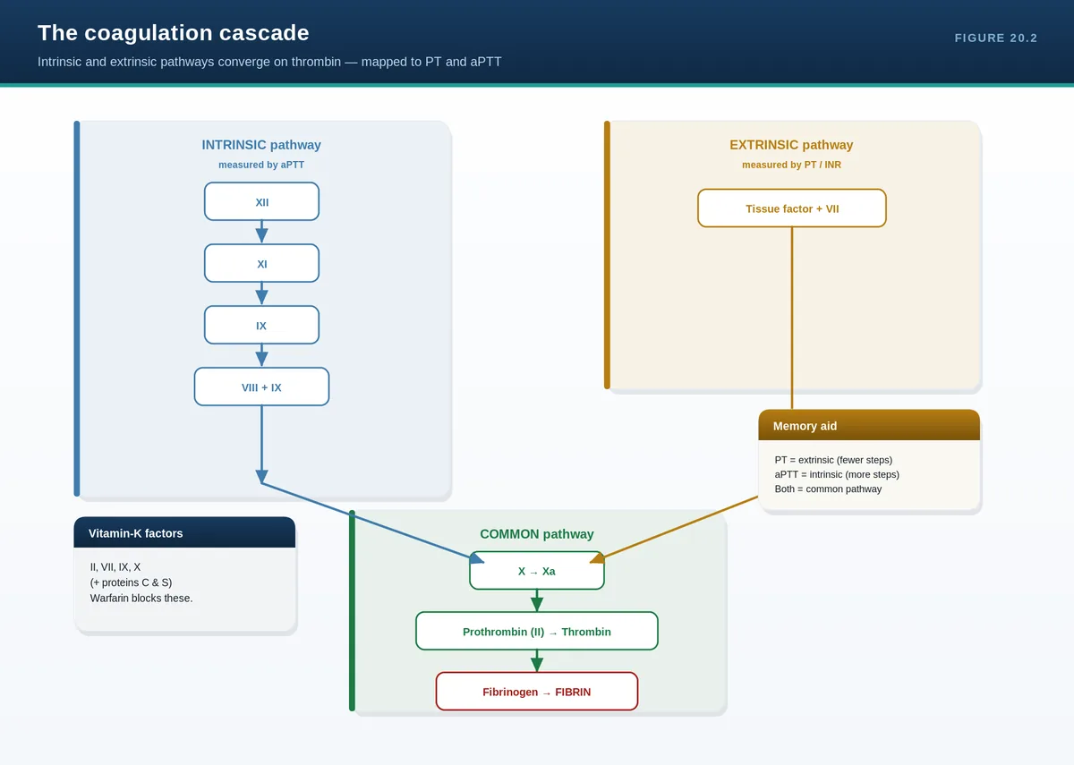
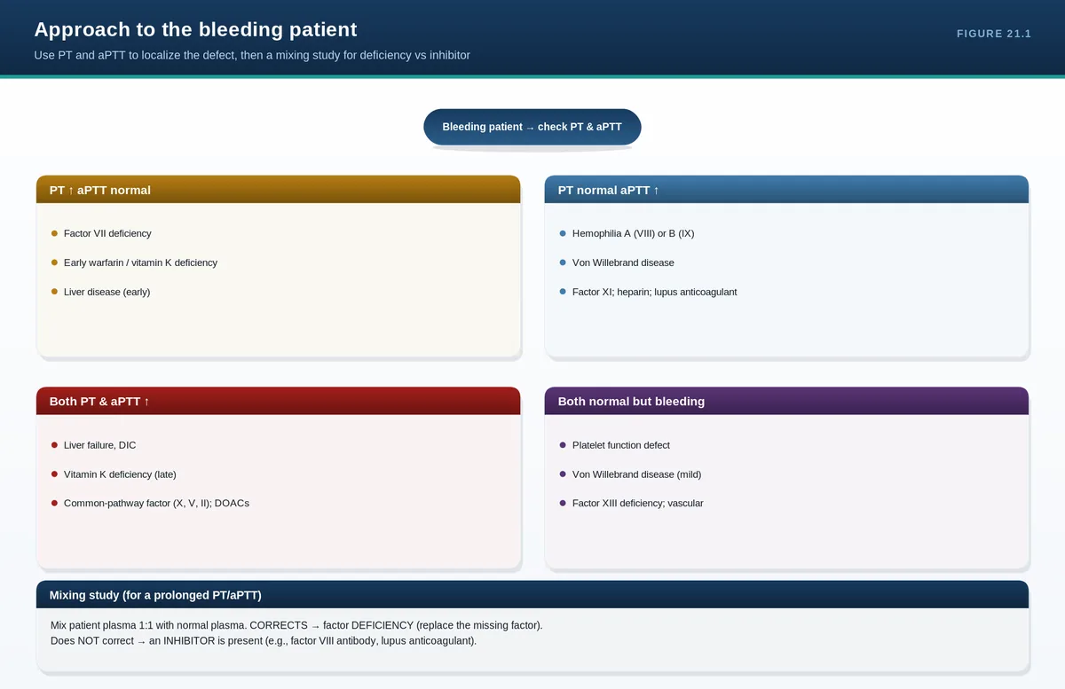
Two tests map the clotting cascade. The PT (reported as INR) tests the extrinsic and common pathways—factor VII and below; it is prolonged by warfarin, liver disease, and vitamin K deficiency. The aPTT tests the intrinsic and common pathways—factors VIII, IX, XI, XII; it is prolonged by heparin, hemophilia, and von Willebrand disease. When a test is prolonged, the mixing study tells you why: mix patient plasma 1:1 with normal plasma. If it corrects, a factor is deficient (replace the missing factor). If it does not correct, an inhibitor is present (e.g., a factor inhibitor or lupus anticoagulant).
Pattern
Likely cause
↑ PT, normal aPTT
Factor VII deficiency, early warfarin, early liver disease, vitamin K deficiency
Normal PT, ↑ aPTT
Hemophilia A/B, von Willebrand disease, heparin, lupus anticoagulant
↑ PT and ↑ aPTT
Liver disease, DIC, vitamin K deficiency, common-pathway factor deficiency
Both normal but bleeding
Platelet disorder, von Willebrand disease, factor XIII deficiency, vascular cause
PITFALLS & PEARLS
Low ferritin = iron deficiency, full stop. But normal/high ferritin does NOT exclude it when inflammation is present—check transferrin saturation.
Haptoglobin is the most sensitive single marker of intravascular hemolysis (it drops first).
A positive direct antiglobulin (Coombs) test means immune-mediated hemolysis; a negative test pushes you toward membrane, enzyme, or mechanical causes.
Mixing study: corrects = factor deficiency; doesn’t correct = inhibitor. This one rule directs the entire bleeding workup.
Check iron studies before transfusing or starting iron when the diagnosis is unclear—transfusion and treatment alter the results.
CASE VIGNETTE
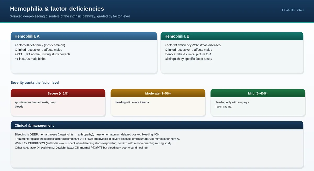
A 6-year-old boy bleeds excessively after a tooth extraction. PT is normal, aPTT is prolonged, and a 1:1 mixing study corrects the aPTT.
Reasoning: isolated aPTT prolongation that corrects on mixing indicates a factor deficiency in the intrinsic pathway—factor VIII or IX assays will distinguish hemophilia A from B.
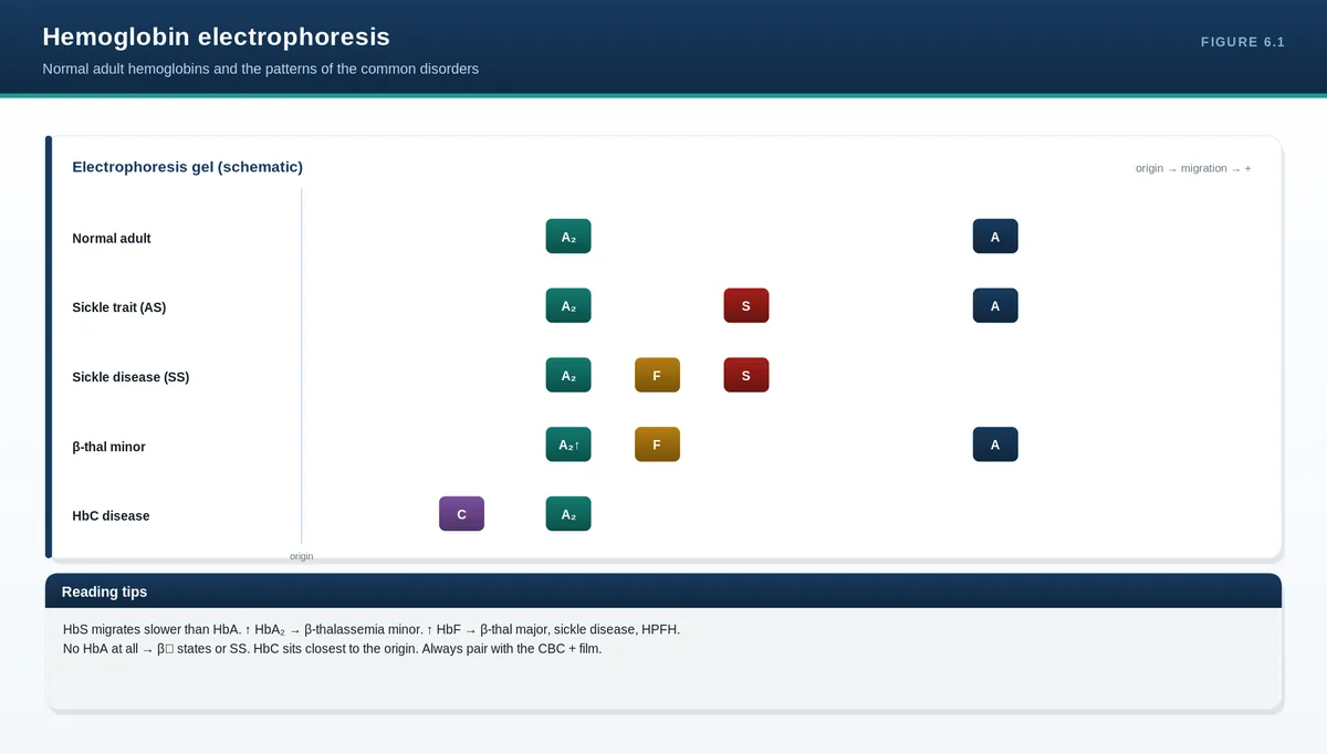
Hemoglobin electrophoresis answers one question: which hemoglobins are present, and in what proportions? Read alongside the CBC, it separates two fundamentally different problems—making too little normal hemoglobin (thalassemia, a quantitative defect) versus making an abnormal hemoglobin (a variant such as HbS, a qualitative defect). A useful analogy: the CBC tells you how much milk is in the carton (the hemoglobin concentration); electrophoresis tells you what kind of milk it is (the mix of hemoglobin types).
Normal hemoglobins and the developmental switch
Hemoglobin is a tetramer: a heme group (iron in protoporphyrin) bound within each of four globin chains. Each red cell carries millions of hemoglobin molecules. Which chains are made changes across life—erythroblasts make a roughly equivalent amount of alpha chains and of the partner chains (beta, delta, gamma), and the partner that dominates shifts with development.
Hemoglobin
Chains
Typical adult %
Note
HbA
α2 β2
~97%
The main adult hemoglobin
HbA2
α2 δ2
2.5–3.5%
Rises in beta-thalassemia trait
HbF
α2 γ2
<1%
Dominant in the fetus; falls after birth
The fetus makes mainly HbF, which then switches to HbA over the first months of life and is nearly complete by about six months. This switch has a key clinical consequence: beta-chain disorders (beta-thalassemia, sickle cell disease) are not apparent at birth because gamma chains are still being made, and only declare themselves after the switch. Alpha-chain disorders, by contrast, affect every hemoglobin (all use alpha chains) and can be detected at birth.
How hemoglobins are separated
Several methods separate hemoglobins, mostly by electrical charge. Traditional gel electrophoresis runs the sample at alkaline (and, to resolve ambiguities, acid) pH. High-performance liquid chromatography (HPLC) is now the standard quantitative method, precisely measuring HbA, HbA2, HbF, and variants. Isoelectric focusing and capillary electrophoresis are also used. The aim in every case is the same: separate the hemoglobin types to diagnose a hemoglobinopathy or thalassemia.
Charge-based methods have a limitation: some variants co-migrate (for example HbS can run with HbD or HbG on an alkaline gel). When a band is ambiguous, confirm it with a second method—an acid gel, HPLC, or a sickle solubility test for HbS.
PITFALLS & PEARLS
Always read the CBC and film BEFORE interpreting electrophoresis—microcytosis, the red cell count, and the clinical picture reframe every result.
Charge-based separation can make different variants co-migrate; confirm an ambiguous band with a second method (acid gel, HPLC, or sickle solubility).
Quantitative versus qualitative — the core distinction
This is the conceptual heart of the chapter. In thalassemia (quantitative), production of a normal chain is reduced, so the hemoglobin concentration is low and the cells are microcytic, but the mix of hemoglobin types is broadly preserved (with the compensatory shifts described below). In a structural variant such as sickle cell disease (qualitative), the problem is not how much hemoglobin is made but that an abnormal hemoglobin (HbS) is produced—so a new band appears on electrophoresis. Returning to the analogy: thalassemia is too little milk of roughly the normal kind; a variant is a different kind of milk in the carton.
Beta-thalassemia patterns
In beta-thalassemia, beta-chain output is reduced, so unpartnered alpha chains pair instead with delta chains (raising HbA2) and with gamma chains (raising HbF). A raised HbA2 above 3.5% is the hallmark of beta-thalassemia trait. The severity spectrum runs from minor (trait) through intermedia to major. Remember the developmental switch: at birth a beta-thalassemia major patient can have near-normal indices because gamma chains still predominate, and the disease emerges only as HbF falls.
Condition
HbA
HbA2
HbF
Hb (g/dL)
MCV
Normal
~95%
2.5%
~1%
12–18
80–100
Beta-thal minor (trait)
90+%
>3.5%
variable
10–12
Low
Beta-thal intermedia
90+%
>3.5%
variable
8–10
Low
Beta-thal major (treated)
90+% transfusion-derived
>3.5%
high
maintained by transfusion
normal on transfusion
Alpha-thalassemia patterns
Alpha-thalassemia is different because alpha chains are needed for HbA, HbA2, and HbF alike—so reduced alpha output cannot raise A2 or F. The electrophoresis is therefore often normal or shows low-normal HbA2 and HbF, and the diagnosis is frequently one of exclusion (microcytosis with normal iron and a normal or low-A2 pattern), confirmed by genetic testing. When alpha output is severely reduced, excess beta chains pair with each other to form HbH (beta-tetramers), seen in three-gene deletion alpha-thalassemia and accompanied by golf-ball inclusions on a supravital-stained film; in the fetus, excess gamma chains form Hb Barts (gamma-tetramers).
PITFALLS & PEARLS
Alpha-thalassemia typically gives a NORMAL or low-A2 electrophoresis—do not expect a raised band; diagnosis is often clinical and genetic.
HbH (β4) with golf-ball red cell inclusions indicates clinically significant (≥3 gene) alpha-thalassemia.
Sickle cell syndromes
Sickle cell disorders are defined by the presence of HbS and by how much normal HbA accompanies it. The single most useful teaching point: in sickle cell trait (HbAS) there is MORE HbA than HbS, whereas in sickle cell disease (HbSS or sickle-beta-0) there is NO HbA at all. Sickle-beta-0-thalassemia behaves like HbSS clinically but adds microcytosis and a raised HbA2/HbF; sickle-beta-plus retains some HbA (but less than 50%) and is usually milder.
Genotype
Hb (g/dL)
HbA
HbS
HbA2 / HbF
Normal
12–15
~95%
0
normal
Sickle trait (HbAS)
12–15
~60% (more A)
~40% (less S)
normal
Sickle disease (HbSS)
6–10
0
90+%
normal
Sickle-beta-0 thal
6–10
0
90+%
↑ A2 ± ↑ F (microcytic)
Sickle-beta+ thal
6–11
variable (<50%)
60+%
↑ A2 ± ↑ F (microcytic)
Compound heterozygotes and genetic counseling
Inheriting two different abnormal beta genes produces a compound heterozygous state. Sickle-beta-0-thalassemia is the classic example—one sickle gene and one beta-0 gene give no HbA and a phenotype like HbSS. This is why partner testing matters: a mother with sickle trait (HbAS) and a father who is a beta-0-thalassemia carrier can, in a quarter of pregnancies, have a child with sickle-beta-0-thalassemia—clinically significant sickle cell disease. HbSC disease (one sickle, one HbC gene) is another important compound state. Counseling depends on identifying both parents' genotypes.
Interpretation at a glance
Electrophoresis finding
Interpretation
↑ HbA2 (>3.5%) ± ↑ HbF, with microcytosis
Beta-thalassemia (trait / intermedia / major or compound state)
Normal or low HbA2/HbF, with microcytosis
Alpha-thalassemia (diagnosis often by exclusion ± genetics)
HbH band (± golf-ball cells)
Alpha-thalassemia with ≥3 gene deletion (excess beta-tetramers)
HbS present WITH HbA (A usually <40%)
Sickle trait (HbAS) or sickle-beta-plus thalassemia
HbS present, NO HbA, ± ↑ A2/F
Sickle cell disease: HbSS, or sickle-beta-0 (if ↑A2/F + microcytic)
PITFALLS & PEARLS
HbA2 >3.5% = beta-thalassemia trait—but coexisting IRON DEFICIENCY lowers HbA2 and can mask it; treat the iron deficiency and repeat the test.
Recent transfusion adds donor HbA and distorts the picture—wait about three months before interpreting.
Hydroxyurea raises HbF; account for it when reading a sickle patient's result.
Before six months of age HbF is physiologically high, so beta disorders can be missed—alpha disorders and Hb Barts, however, are detectable at birth.
Sickle trait (AS) has MORE A than S; sickle disease (SS / Sβ0) has NO A. This one rule separates carrier from patient.
CASE VIGNETTE
A 54-year-old woman has a long-standing history of microcytic anemia and no chronic illness. Iron studies are normal. Hemoglobin electrophoresis shows HbA2 of 5%.
Reasoning: persistent microcytic anemia with NORMAL iron is not iron deficiency—an electrophoresis showing HbA2 >3.5% confirms beta-thalassemia trait. The lesson is not to keep treating "refractory iron deficiency" with iron; check the electrophoresis (and, in younger patients, counsel about inheritance).
QUICK REVIEW
Electrophoresis = which hemoglobins are present and how much; always read with the CBC.
Normal: HbA (α2β2) ~97%, HbA2 (α2δ2) 2.5–3.5%, HbF (α2γ2) <1%; HbF→HbA switch by ~6 months.
Quantitative (thalassemia, less hemoglobin) vs qualitative (variant, abnormal hemoglobin) — milk amount vs milk type.
↑ HbA2 (>3.5%) ± ↑ HbF = beta-thal trait; iron deficiency lowers HbA2 (retest after iron).
Alpha-thal: normal/low pattern ± HbH. Sickle: AS has more A than S; SS / Sβ0 has no A.
Most of hematology can be navigated with three algorithms and one safety list. Anemia is solved with size and reticulocytes. Bleeding is solved by deciding primary versus secondary hemostasis. Abnormal counts are solved by asking reactive versus clonal. And a short list of emergencies must be recognized on sight. These pathways tie Part I together and preview the disease chapters to come.
Algorithm 1 — Approach to anemia
Confirm anemia and check it is real (not dilutional or a lab error).
Classify by MCV: microcytic, normocytic, or macrocytic.
Check the reticulocyte count: high (bleeding/hemolysis) vs low (production problem).
Direct testing by the box you land in: microcytic → iron studies (± electrophoresis); macrocytic → B12/folate, TSH, liver, film; normocytic with high retics → hemolysis panel ± film; normocytic with low retics → renal function, marrow assessment.
Always ask “why”: even when you find the type, identify the underlying cause (e.g., the source of blood loss).
Algorithm 2 — Approach to the bleeding patient
Characterize the bleeding: mucocutaneous (petechiae, bruising, epistaxis, menorrhagia) suggests primary hemostasis; deep bleeds into joints/muscles suggest secondary hemostasis.
Screen with platelet count, PT, and aPTT; add a film.
Low platelets → is it production, destruction (e.g., ITP), or sequestration? Confirm it’s not clumping.
Normal platelets but mucocutaneous bleeding → think von Willebrand disease or a platelet function defect.
Abnormal PT/aPTT → use the mixing study to separate deficiency from inhibitor; map the pattern to the cascade.
Algorithm 3 — Approach to abnormal counts (high or low)
For any abnormal line, first ask: reactive (secondary) or clonal (primary)?
Reactive clues: an obvious trigger (infection, inflammation, iron deficiency, hypoxia, steroids), proportionate changes, and a normal film.
Clonal clues: very high or persistent counts, multiple lines involved, abnormal/immature cells on the film, splenomegaly, or constitutional symptoms.
Cytopenias: decide one line versus pancytopenia—pancytopenia raises marrow failure, infiltration, megaloblastic anemia, or hypersplenism.
When clonal disease is suspected, the bone marrow examination and cytogenetics become the key next step.
Hematologic emergencies — recognize on sight
Emergency
Trigger to recognize
Immediate concern
Thrombotic thrombocytopenic purpura (TTP)
Schistocytes + low platelets
Start urgent plasma exchange; do not just transfuse platelets
Acute leukemia / blasts
Blasts or Auer rods on film, pancytopenia
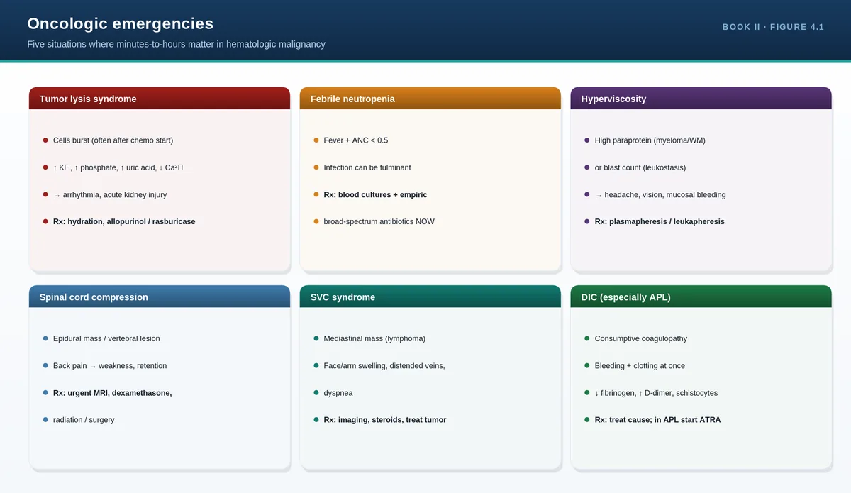
Urgent hematology referral; watch for tumor lysis, DIC
Febrile neutropenia
Fever with neutrophils <0.5 ×10⁹/L
Immediate broad-spectrum antibiotics
Disseminated intravascular coagulation (DIC)
Bleeding + ↑PT/aPTT, ↓platelets, ↓fibrinogen
Treat the trigger; support with products
Severe symptomatic anemia
Hb very low with cardiac compromise
Transfuse; find and stop the cause
PITFALLS & PEARLS
When two algorithms collide, safety first: schistocytes + low platelets is treated as TTP until excluded, whatever else is going on.
“Reactive vs clonal” is the question behind every abnormal count—write it at the top of your differential.
Don’t stop at the label: “iron deficiency anemia” is a finding, not a diagnosis—the diagnosis is the cause of the iron loss.
Pancytopenia always earns a film and usually a marrow examination.
CASE VIGNETTE
A 70-year-old man has Hb 7 g/dL, WBC 2.0 ×10⁹/L, platelets 40 ×10⁹/L, and the film shows circulating blasts.
Reasoning: pancytopenia with blasts is acute leukemia until proven otherwise—an emergency. The algorithms converge: abnormal counts (clonal), and an emergency pattern (blasts) that mandates urgent referral and marrow examination.
QUICK REVIEW
Anemia: size (MCV) then response (reticulocytes), then target testing, then find the cause.
Bleeding: primary (mucocutaneous) vs secondary (deep); screen with platelets, PT, aPTT, film.
Abnormal counts: reactive vs clonal; pancytopenia → film ± marrow.
Emergencies to know on sight: TTP, acute leukemia/blasts, febrile neutropenia, DIC, severe anemia.
A label is not a diagnosis—always ask why.
With these foundations in place—the lineage map, cell physiology, the CBC, the blood film, the core laboratory panels, and the master algorithms—the disease chapters become applications of a framework you already know. Part II turns to the anemias, the largest and most clinically common group, where the two-number method from Chapter 3 does most of the diagnostic work.
Part II applies the framework from Part I to the anemias—the largest and most common group of hematologic problems you will meet. Each chapter keeps the same structure (the big idea, then pathophysiology, clinical clues, diagnosis and management, pitfalls and pearls, a case, and a quick review), and leans on the two-number method—red cell size and reticulocyte response—introduced in Chapter 3. The chapters are numbered continuously from Part I, beginning here at Chapter 8.
Anemia is a sign, not a diagnosis. The job is always two-fold: classify the anemia (so the differential shrinks) and find its cause (so you actually treat the patient). Two numbers do most of the classifying—the MCV tells you cell size, the reticulocyte count tells you whether the marrow is responding—and the cause is then pursued with targeted tests and a careful history.
Definition and physiology
Anemia is a reduced hemoglobin concentration—below about 13 g/dL in men and 12 g/dL in non-pregnant women—lowering the blood's oxygen-carrying capacity. Symptoms (fatigue, breathlessness, pallor, palpitations) come from reduced tissue oxygen delivery and from compensation: the heart rate and cardiac output rise, 2,3-BPG increases to unload more oxygen, and erythropoietin drives the marrow. How sick a patient feels depends less on the absolute hemoglobin than on how quickly it fell: a slow drop to 7 g/dL may be well tolerated, while an acute drop to 9 g/dL can cause collapse.
Two ways to classify
The first axis is cell size (MCV); the second is the underlying mechanism—reduced production, increased destruction (hemolysis), or blood loss. The reticulocyte count links the two: it is high when cells are being lost or destroyed and low when production fails.
MCV category
Common causes
Microcytic (<80 fL)
Iron deficiency, thalassemia, anemia of chronic disease (some), sideroblastic
History targets the cause: blood loss (menstrual, gastrointestinal, including dark stools), diet (vegan, alcohol), drugs, family history (hemoglobinopathy, gallstones, splenectomy), and chronic disease. Examination looks for pallor, jaundice and splenomegaly (hemolysis), koilonychia and glossitis (iron and B12), and lymphadenopathy. Then the labs flow logically: CBC and film, classify by MCV, check reticulocytes, and order targeted tests—iron studies for microcytosis, B12/folate for macrocytosis, a hemolysis panel when reticulocytes are high.
PITFALLS & PEARLS
Anemia is a finding, not a final answer—"iron deficiency anemia" still requires you to find the source of iron loss.
Rate of fall matters more than the number: a chronic Hb of 7 may walk in; an acute Hb of 9 may be in shock.
A low reticulocyte count in the face of anemia is inappropriate—it tells you production has failed.
Mixed pictures (e.g., iron + B12 deficiency) can give a normal MCV with a high RDW—let the film and RDW flag them.
CASE VIGNETTE
A 68-year-old man has fatigue, Hb 8 g/dL, MCV 74 fL, and a high RDW; reticulocytes are low.
Reasoning: microcytic anemia with low reticulocytes is a production problem—iron deficiency tops the list. In an older man without obvious bleeding, the cause must be sought in the gastrointestinal tract (occult malignancy) before simply prescribing iron.
QUICK REVIEW
Classify by MCV (size) and reticulocytes (response); then find the cause.
Micro: iron, thalassemia; normo: bleeding/hemolysis/chronic; macro: B12/folate and others.
High retic = loss/destruction; low retic = production failure.
History (blood loss, diet, drugs, family) + exam (pallor, jaundice, spleen) guide targeted tests.
Always identify the underlying cause, not just the anemia type.
Iron deficiency is the most common anemia worldwide, but it is never the whole answer—it is a clue pointing to blood loss, increased demand, or malabsorption. The body has no way to excrete iron, so balance is controlled entirely at absorption, governed by the hormone hepcidin. Understand hepcidin and both deficiency and overload make sense.
Iron physiology and hepcidin
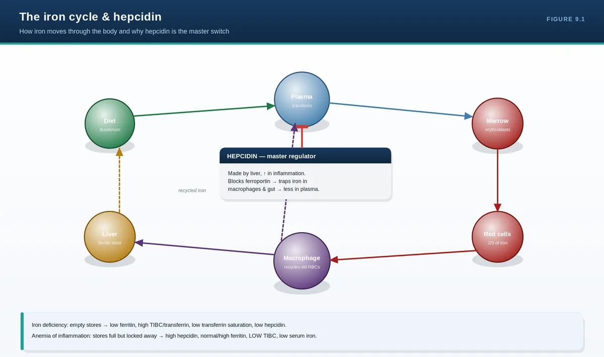
Dietary iron is absorbed in the duodenum, carried in plasma bound to transferrin, used by erythroid precursors to make hemoglobin, and stored as ferritin. Senescent red cells are broken down by macrophages, which recycle their iron back to plasma—this recycling supplies most daily needs. Hepcidin, made by the liver, is the master regulator: it lowers iron absorption from the gut and blocks release from macrophages by degrading the iron exporter ferroportin. Hepcidin falls in iron deficiency (letting more iron in) and rises in inflammation (trapping iron away), which is the link to anemia of chronic disease.
Causes—and why the cause matters
Blood loss (most important): menstrual loss in younger women; gastrointestinal loss (ulcer, malignancy) in men and postmenopausal women—this group needs GI evaluation.
Increased demand: pregnancy, infancy, and rapid growth.
Malabsorption: celiac disease, gastrectomy, bariatric surgery, H. pylori.
Inadequate intake: poor diet, contributing rather than acting alone in adults.
Clinical and laboratory features
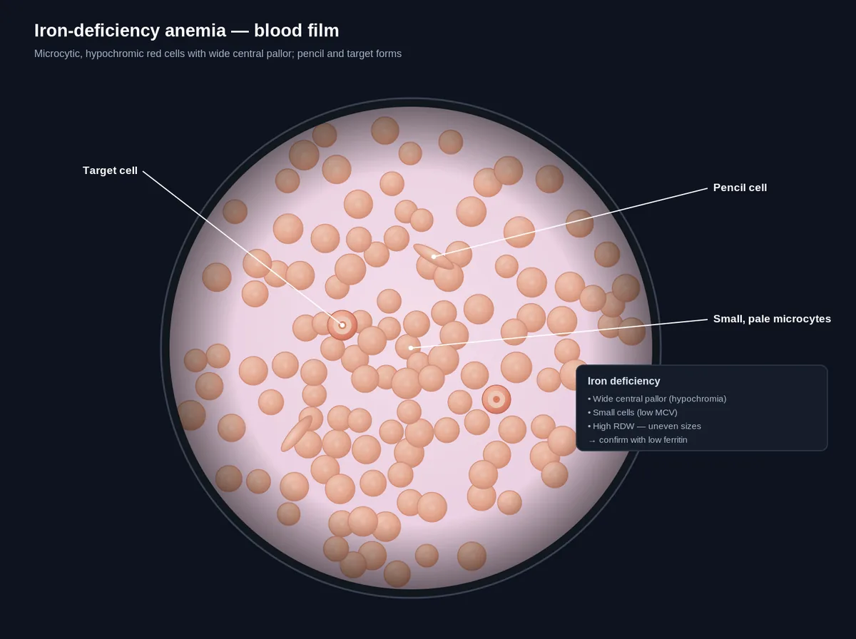
Beyond general anemia symptoms, iron deficiency produces koilonychia (spoon nails), glossitis, angular cheilitis, pica (craving ice or non-foods), and restless legs. The blood picture is microcytic and hypochromic with a high RDW and often reactive thrombocytosis. Iron studies show low ferritin (the most specific test), low serum iron, high transferrin/TIBC, and low transferrin saturation—remembering that ferritin can be falsely normal or high with coexisting inflammation.
Management
Treat the cause and replace iron. Oral iron is first line; alternate-day dosing improves absorption and tolerability. The expected response is a reticulocyte rise within a week and a hemoglobin rise of about 2 g/dL over three to four weeks—then continue for several months to refill stores. Intravenous iron is used for intolerance, malabsorption, ongoing heavy loss, chronic kidney disease, or when rapid repletion is needed. Failure to respond should prompt rechecking adherence, absorption, and ongoing blood loss.
Iron overload
Too much iron is also harmful. Hereditary hemochromatosis (commonly HFE mutations) causes excess absorption; transfusional overload accumulates in chronically transfused patients (e.g., thalassemia). Excess iron deposits in the liver (cirrhosis), heart (cardiomyopathy, arrhythmia), pancreas and endocrine organs (diabetes, hypogonadism), skin (bronze pigmentation), and joints. Ferritin and transferrin saturation are high. Treatment is therapeutic phlebotomy for hemochromatosis and iron chelation for transfusional overload.
PITFALLS & PEARLS
Iron deficiency in a man or postmenopausal woman is gastrointestinal cancer until proven otherwise—investigate the gut.
Low ferritin confirms iron deficiency; a normal/high ferritin does not exclude it when inflammation is present (check transferrin saturation).
Alternate-day oral iron is absorbed better than multiple daily doses (lower hepcidin between doses).
Reticulocytosis within days is the first sign oral iron is working—before the hemoglobin moves.
Do not phlebotomize an iron-overloaded patient who is anemic from transfusions—use chelation instead.
CASE VIGNETTE
A 70-year-old man has iron-deficiency anemia and no overt bleeding. Colonoscopy reveals a right-sided colon cancer.
Reasoning: occult gastrointestinal bleeding is the leading cause of iron deficiency in older men. Prescribing iron without investigating would have missed a curable malignancy—find the cause first.
QUICK REVIEW
Iron: absorbed in duodenum, carried on transferrin, stored as ferritin, recycled by macrophages; no excretion route.
Hepcidin is the master regulator (low in deficiency, high in inflammation).
IDA = microcytic/hypochromic, low ferritin, low sat, high TIBC, high RDW; find the cause (often GI loss).
Treat: oral iron (alternate-day), IV iron when indicated; continue to replete stores.
Overload (hemochromatosis/transfusional): high ferritin & sat, organ damage; treat by phlebotomy or chelation.
The iron cycle & hepcidinIron studies — pattern recognitionIron-deficiency anemia — blood film
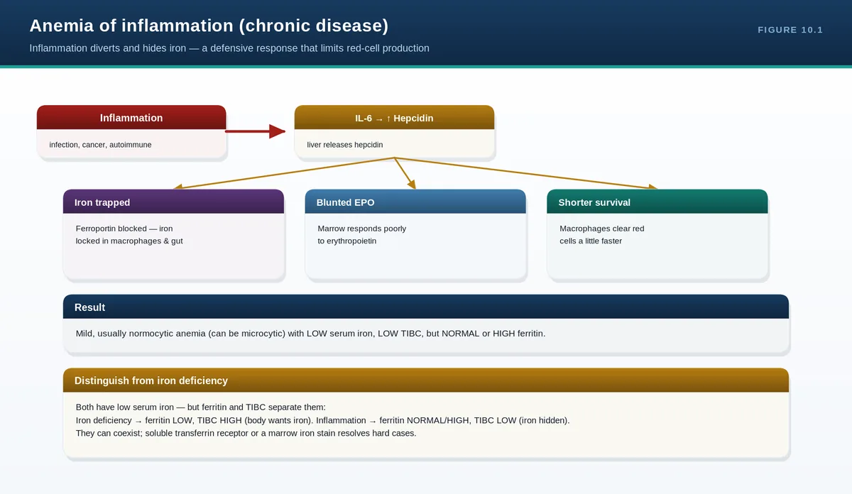
Chronic inflammation tells the body to hide its iron. High hepcidin locks iron inside macrophages, erythropoietin signaling is blunted, and red cell survival shortens—producing a usually mild, normocytic anemia despite adequate (even increased) iron stores. It is a functional iron deficiency: plenty of iron, but none available to the marrow.
Pathogenesis
Inflammatory cytokines, especially interleukin-6, drive the liver to make hepcidin. Hepcidin traps iron in macrophages and reduces gut absorption, so serum iron falls even though stores (ferritin) are normal or high. At the same time, cytokines blunt the marrow's response to erythropoietin and modestly shorten red cell lifespan. The result is the anemia seen in chronic infections, autoimmune disease, and malignancy. The most effective treatment is to control the underlying disease.
Telling it apart from iron deficiency
Test
Iron deficiency
Anemia of inflammation
Ferritin
Low
Normal or high
Serum iron
Low
Low
TIBC/transferrin
High
Low or normal
Transferrin saturation
Low
Low or normal
Soluble transferrin receptor
High
Normal
The two can coexist—a high ferritin from inflammation can mask true iron deficiency. A low transferrin saturation with a normal-range ferritin, or a high soluble transferrin receptor, helps unmask combined deficiency.
Anemia of renal failure
Chronic kidney disease causes anemia chiefly through reduced erythropoietin production by the failing kidney (with contributions from uremia and inflammation). It is treated with erythropoiesis-stimulating agents and iron, targeting a modest hemoglobin (around 10–11 g/dL)—not normal levels, because over-correction raises the risk of thrombosis and cardiovascular events.
PITFALLS & PEARLS
Anemia of inflammation is functional iron deficiency: iron is present but locked away by hepcidin.
It is usually mild and normocytic; a severe or very microcytic anemia should make you look for a second cause.
A normal ferritin in a sick, inflamed patient does not exclude true iron deficiency—check saturation or sTfR.
In renal anemia, aim for a modest hemoglobin target; full normalization with ESAs increases cardiovascular risk.
CASE VIGNETTE
A patient with rheumatoid arthritis has Hb 10.5 g/dL, MCV 88 fL, low serum iron, low TIBC, and a high-normal ferritin.
Reasoning: the low TIBC and preserved ferritin distinguish anemia of inflammation from iron deficiency (where TIBC is high and ferritin low). Treatment is better disease control, not reflexive iron.
QUICK REVIEW
Cytokines (IL-6) → hepcidin → iron trapped in macrophages; blunted EPO; shortened RBC survival.
Usually mild, normocytic; ferritin normal/high, TIBC low (opposite of IDA).
sTfR and saturation help unmask coexisting iron deficiency.
Renal anemia = EPO deficiency; treat with ESA + iron to a modest target.
Best treatment is controlling the underlying disease.
B12 and folate are needed to make DNA. Without them, cells cannot divide properly even though the cytoplasm keeps maturing—producing large, abnormal cells, ineffective erythropoiesis, and eventually pancytopenia. The clinical trap is unique to B12: its deficiency also damages the nervous system, often irreversibly, so you must never treat blindly with folate alone.
Biochemistry and absorption
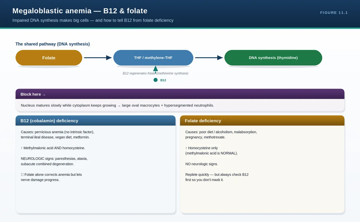
Folate and B12 cooperate in DNA synthesis: folate provides one-carbon units for making thymidine, and B12 is required to regenerate the active form of folate (via the methionine synthase reaction). When either is missing, DNA replication stalls while RNA and protein synthesis continue, causing nuclear–cytoplasmic asynchrony—the megaloblastic change. B12 also runs a separate reaction (methylmalonyl-CoA mutase) important for myelin, which is why its deficiency causes neurological disease. Absorption differs: B12 needs intrinsic factor from gastric parietal cells and is taken up in the terminal ileum; folate is absorbed in the jejunum and has small body stores (deficiency develops in months), whereas B12 stores last years.
Pernicious anemia—autoimmune destruction of parietal cells with anti-intrinsic-factor antibodies—is the classic cause of B12 deficiency and is associated with other autoimmune disease and a long-term gastric cancer risk.
Clinical and laboratory features
Both cause a macrocytic anemia with glossitis, mild jaundice (from ineffective erythropoiesis raising bilirubin and LDH), and, when severe, pancytopenia. B12 deficiency adds neurological features: a peripheral neuropathy, subacute combined degeneration of the cord (dorsal columns—proprioception and vibration loss), and cognitive or psychiatric changes. The blood film shows oval macrocytes and hypersegmented neutrophils (six or more lobes), which can appear before the MCV rises. Confirmatory tests include low B12 or folate, and raised methylmalonic acid (B12-specific) and homocysteine.
Management
Replace the deficient vitamin—B12 by injection (or high-dose oral) and folate orally. The cardinal rule: in combined or unclear deficiency, give B12 before or together with folate, because folate alone can correct the anemia while allowing the neurological damage of B12 deficiency to progress. Watch for hypokalemia as the marrow regenerates and consumes potassium during early treatment.
PITFALLS & PEARLS
Never treat megaloblastic anemia with folate alone until B12 deficiency is excluded—you can precipitate or worsen irreversible neurological damage.
Hypersegmented neutrophils are an early, high-yield clue—often present before the MCV is clearly raised.
Methylmalonic acid is elevated in B12 deficiency but not folate deficiency—use it to distinguish them.
B12 stores last years; folate stores last only months—folate deficiency develops far more quickly.
Watch for hypokalemia in the first days of treatment as new cells take up potassium.
CASE VIGNETTE
A 60-year-old vegetarian has Hb 9 g/dL, MCV 116 fL, hypersegmented neutrophils, mild jaundice, and numb, tingling feet with reduced vibration sense.
Reasoning: macrocytic anemia with neurological signs points to B12 deficiency (here likely dietary ± pernicious anemia). Confirm with B12 and methylmalonic acid, check anti-IF antibodies, and replace B12—do not give folate alone.
QUICK REVIEW
B12 and folate are needed for DNA synthesis; deficiency → megaloblastic change, ineffective erythropoiesis, pancytopenia.
B12 needs intrinsic factor + terminal ileum; pernicious anemia is the classic cause. Folate: jejunum, short stores.
Film: oval macrocytes + hypersegmented neutrophils; ↑ MMA (B12-specific) and homocysteine.
B12 deficiency causes subacute combined degeneration—neurology may be irreversible.
Replace B12 before/with folate; watch potassium on recovery.
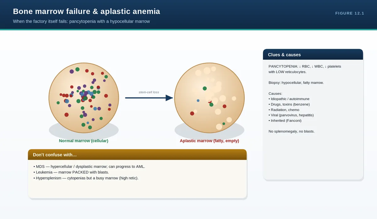
When several cell lines fall together (pancytopenia), suspect the factory. The marrow may be empty (aplastic anemia—immune destruction of stem cells), replaced (infiltration or fibrosis), or dysplastic (myelodysplasia). A bone marrow examination is what separates these, and it changes everything about management.
Approaching pancytopenia
Pancytopenia has two broad mechanisms. Reduced production includes aplastic anemia, myelodysplastic syndromes, marrow infiltration (leukemia, lymphoma, metastatic cancer, fibrosis), and severe megaloblastic anemia. Increased peripheral removal or pooling includes hypersplenism and immune destruction. The reticulocyte count and the bone marrow aspirate and trephine biopsy distinguish an empty marrow (aplastic) from a packed or dysplastic one.
Aplastic anemia
Aplastic anemia is pancytopenia with a hypocellular marrow and no abnormal infiltrate. Most cases are acquired and immune-mediated—cytotoxic T cells attack hematopoietic stem cells. Triggers include drugs (e.g., chloramphenicol, certain anticonvulsants), toxins (benzene), viruses (including seronegative hepatitis), and radiation; many are idiopathic. Inherited marrow failure syndromes (such as Fanconi anemia) present in younger patients and may have congenital anomalies. Clinically, patients show the consequences of each missing line: anemia, infections (neutropenia), and bleeding (thrombocytopenia).
Management
Care has two arms: support and definitive treatment. Support means transfusions (used judiciously), prompt treatment of infection, and removing any offending agent. Definitive treatment depends on age and severity: a young patient with severe disease and a matched sibling donor is offered hematopoietic stem cell transplantation, while others receive immunosuppressive therapy (antithymocyte globulin plus cyclosporine), often with a thrombopoietin agonist. Pure red cell aplasia (an isolated failure of red cell production, linked to parvovirus B19, thymoma, and drugs) is a related but distinct entity.
PITFALLS & PEARLS
Pancytopenia always deserves a blood film and usually a marrow biopsy—do not anchor on one explanation.
Aplastic anemia = hypocellular marrow with NO infiltrate; a packed marrow points to leukemia, MDS, or infiltration instead.
Always take a drug and toxin history—removing the cause can be curative.
In a young patient with severe aplastic anemia, a matched sibling transplant is potentially curative; otherwise immunosuppression.
Parvovirus B19 causes pure red cell aplasia and dangerous aplastic crises in patients with chronic hemolysis.
CASE VIGNETTE
A 22-year-old develops fatigue, bruising, and mouth infections. CBC shows pancytopenia with very low reticulocytes; marrow biopsy is markedly hypocellular with no blasts or infiltrate.
Reasoning: hypocellular marrow with pancytopenia and no infiltrate is aplastic anemia. Management is supportive care plus definitive therapy—transplant if a young patient has a matched donor, otherwise immunosuppression.
QUICK REVIEW
Pancytopenia: production failure (aplastic, MDS, infiltration, megaloblastic) vs peripheral (hypersplenism, immune).
Aplastic anemia = hypocellular marrow, no infiltrate; mostly immune-mediated stem cell destruction.
Causes: drugs, toxins (benzene), viruses, radiation, inherited (Fanconi), idiopathic.
Treat: remove cause + support; HSCT (young, severe, donor) or immunosuppression (ATG + cyclosporine).
Parvovirus B19 → pure red cell aplasia / aplastic crisis.
Bone marrow failure & aplastic anemia
13Hemolytic Anemias: Overview and Proving Hemolysistop ↑
THE BIG IDEA
Hemolysis means red cells are destroyed faster than their normal 120-day lifespan, and the marrow ramps up to compensate (reticulocytosis). The diagnostic flow is always the same: first prove hemolysis is happening, then localize it—inherited or acquired, intravascular or extravascular, immune or non-immune. One test, the direct antiglobulin (Coombs) test, is the pivot between immune and non-immune causes.
Proving hemolysis
Look for evidence of red cell breakdown plus a marrow response. Breakdown raises LDH and unconjugated (indirect) bilirubin and consumes haptoglobin, which falls. The marrow responds with a high reticulocyte count, and the film may reveal the mechanism. Together—high reticulocytes, high LDH, high indirect bilirubin, low haptoglobin—these confirm hemolysis before you ask why.
Localizing: intravascular versus extravascular
Feature
Intravascular
Extravascular
Site
Within blood vessels
Spleen/liver macrophages
Haptoglobin
Very low
Low/normal
Hemoglobinuria
Present
Absent
Film clue
Schistocytes, bite, blister cells
Spherocytes
Examples
MAHA, transfusion reaction, severe G6PD, PNH
Hereditary spherocytosis, warm AIHA
A classification you can reproduce
Inherited hemolysis reflects an intrinsic red cell defect: the membrane (hereditary spherocytosis), an enzyme (G6PD, pyruvate kinase), or the hemoglobin (sickle cell, thalassemia). Acquired hemolysis is usually an extrinsic assault: immune (autoimmune, alloimmune, drug-induced), mechanical (microangiopathy, prosthetic valves), infection (malaria), or the acquired membrane defect of paroxysmal nocturnal hemoglobinuria. After proving hemolysis, the direct antiglobulin test sorts immune (positive) from non-immune (negative) causes and directs the rest of the workup.
PITFALLS & PEARLS
The hemolysis quartet: ↑ reticulocytes, ↑ LDH, ↑ indirect bilirubin, ↓ haptoglobin.
Haptoglobin falls earliest and furthest in intravascular hemolysis; hemoglobinuria signals it too.
The direct antiglobulin (Coombs) test is the hinge: positive = immune; negative = membrane, enzyme, mechanical, or PNH.
Spherocytes = hereditary spherocytosis OR warm autoimmune hemolysis—use the DAT to tell them apart.
CASE VIGNETTE
A patient with jaundice has Hb 8 g/dL, reticulocytes 12%, LDH high, indirect bilirubin high, and haptoglobin undetectable.
Reasoning: the quartet confirms hemolysis. The next steps are a blood film and a direct antiglobulin test to localize and classify—immune versus non-immune—before treating.
QUICK REVIEW
Hemolysis = shortened RBC survival + marrow compensation (reticulocytosis).
Prove it: ↑ retic, ↑ LDH, ↑ indirect bilirubin, ↓ haptoglobin.
Localize: intravascular (schistocytes, hemoglobinuria) vs extravascular (spherocytes, splenomegaly).
Classify: inherited (membrane/enzyme/hemoglobin) vs acquired (immune/mechanical/infection/PNH).
DAT (Coombs) separates immune from non-immune.
14Hereditary Hemolytic Anemias: Membrane and Enzyme Defectstop ↑
THE BIG IDEA
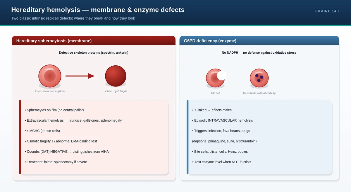
Some red cells are born fragile. A faulty membrane skeleton (hereditary spherocytosis) or a missing protective enzyme (G6PD deficiency) shortens survival. The spleen, the quality-control filter, culls these abnormal cells—so these disorders share splenic destruction, jaundice, and gallstones, but differ sharply in their triggers and blood films.
Hereditary spherocytosis
A defect in membrane-skeleton proteins (commonly spectrin or ankyrin) makes the red cell lose surface area, rounding into a spherocyte. Spherocytes are stiff and are trapped and destroyed in the spleen (extravascular hemolysis). Patients have anemia, jaundice, splenomegaly, and pigment gallstones; severity ranges from mild to transfusion-dependent. The film shows spherocytes, the MCHC is characteristically high, and the direct antiglobulin test is negative (distinguishing it from autoimmune hemolysis). Diagnosis is supported by the EMA-binding test or osmotic fragility. Management is folate supplementation, with splenectomy reserved for more severe disease (after vaccination, given the infection risk).
G6PD deficiency
Glucose-6-phosphate dehydrogenase protects red cells from oxidative stress by generating NADPH. The deficiency is X-linked (affecting mainly but not only males) and common in once malaria-endemic regions. Most of the time patients are well, but exposure to an oxidant—certain drugs (primaquine, dapsone, sulfonamides, nitrofurantoin), fava beans, or infection—triggers acute, often intravascular, hemolysis. The film during a crisis shows bite cells and blister cells, and Heinz bodies (denatured hemoglobin) on supravital staining. Management is to identify and remove the trigger and provide supportive care; enzyme levels should be measured after recovery, since they can be falsely normal during an acute episode when the most deficient cells have already lysed.
Pyruvate kinase deficiency
The most common glycolytic enzyme defect, pyruvate kinase deficiency is autosomal recessive and reduces ATP production, causing a chronic non-spherocytic hemolytic anemia of variable severity, with extravascular hemolysis and splenomegaly.
PITFALLS & PEARLS
Hereditary spherocytosis: spherocytes + high MCHC + NEGATIVE direct antiglobulin test (vs warm AIHA, which is positive).
Vaccinate against encapsulated organisms before any splenectomy, and give folate to all chronic hemolysis patients.
G6PD is X-linked—ask about new drugs, fava beans, or recent infection in a male with acute hemolysis.
A G6PD level can be falsely normal right after a hemolytic crisis (reticulocytes are enzyme-rich)—retest weeks later.
Bite cells and Heinz bodies point to oxidative hemolysis (G6PD).
CASE VIGNETTE
A young man of Mediterranean descent develops dark urine and jaundice days after starting an antimalarial. The film shows bite cells; haptoglobin is undetectable.
Reasoning: drug-induced oxidative, intravascular hemolysis is classic for G6PD deficiency. Stop the trigger and support him; confirm the enzyme level after recovery, not during the crisis.
QUICK REVIEW
Inherited hemolysis from intrinsic defects: membrane (HS), enzyme (G6PD, PK).
HS: spectrin/ankyrin defect → spherocytes, ↑MCHC, negative DAT; folate, splenectomy if severe.
G6PD: X-linked; oxidant triggers (drugs, fava beans, infection) → bite cells, Heinz bodies, intravascular hemolysis.
G6PD level can be falsely normal during a crisis—retest later.
PK deficiency: chronic non-spherocytic hemolytic anemia.
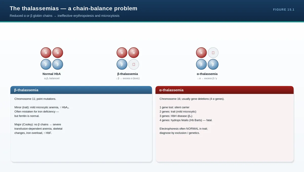
Thalassemia is a problem of quantity, not quality: a globin chain is made in too small an amount. The resulting imbalance—too much of the unaffected chain—damages developing red cells in the marrow (ineffective erythropoiesis) and shortens their survival, giving a microcytic anemia whose severity tracks the genetics. Read this chapter alongside the electrophoresis chapter (Chapter 6).
Genetics and pathophysiology
Hemoglobin needs balanced production of alpha and beta chains. In beta-thalassemia (usually point mutations) beta-chain output is reduced, so excess alpha chains precipitate and damage red cell precursors. In alpha-thalassemia (usually gene deletions—there are four alpha genes) alpha output is reduced, and excess beta or gamma chains form unstable tetramers (HbH or Hb Barts). The shared theme is chain imbalance causing ineffective erythropoiesis and hemolysis.
Beta-thalassemia
Beta-thalassemia minor (trait): one affected gene; mild or no anemia, marked microcytosis with a high red cell count, and raised HbA2—important mainly for genetic counseling.
Beta-thalassemia intermedia: moderate anemia, not regularly transfusion-dependent.
Beta-thalassemia major (Cooley anemia): severe transfusion-dependent anemia from infancy, with marrow expansion (frontal bossing, “hair-on-end” skull), extramedullary hematopoiesis, hepatosplenomegaly, and—from transfusions and increased gut absorption—iron overload.
Alpha-thalassemia
Severity depends on how many of the four alpha genes are lost: one (silent carrier), two (alpha-thalassemia trait, microcytosis), three (HbH disease, a moderate-to-severe hemolytic anemia with beta-tetramers and golf-ball inclusions), and four (Hb Barts, causing hydrops fetalis and death in utero unless treated).
Diagnosis and management
The indices are a strong clue: a low MCV with a high-normal or high red cell count and only mild anemia (a low Mentzer index) suggests thalassemia over iron deficiency. Confirm with hemoglobin electrophoresis/HPLC (raised HbA2 in beta-trait) and, where needed, genetic testing. Management ranges from none (trait, plus counseling) to regular transfusion with iron chelation in major disease, folate supplementation, judicious splenectomy, and curative hematopoietic stem cell transplantation in selected children. A practical pitfall: never mistake thalassemia trait for iron deficiency and treat with long-term iron.
PITFALLS & PEARLS
Microcytosis with a HIGH red cell count and only mild anemia suggests thalassemia trait, not iron deficiency (Mentzer index <13).
Beta-thal trait shows raised HbA2; alpha-thal trait shows a normal/low electrophoresis (diagnosis often genetic).
Do not treat thalassemia trait with iron—iron studies are normal and iron loading can result.
In beta-thalassemia major, iron overload (from transfusion and absorption), not anemia, drives long-term mortality—chelate.
Beta disorders appear after the HbF→HbA switch (months of age); four-gene alpha loss (Hb Barts) is lethal in utero.
CASE VIGNETTE
A 25-year-old has Hb 11 g/dL, MCV 68 fL, a red cell count of 6.0 ×10¹²/L, and normal iron studies. Electrophoresis shows HbA2 4.8%.
Reasoning: mild anemia with marked microcytosis, a high red cell count, normal iron, and raised HbA2 is beta-thalassemia trait. No iron is needed; the key action is genetic counseling.
QUICK REVIEW
Thalassemia = reduced globin chain quantity → chain imbalance → ineffective erythropoiesis + hemolysis; microcytic.
Beta (point mutations): minor (↑HbA2), intermedia, major (transfusion-dependent, marrow expansion, iron overload).
Alpha (deletions of 4 genes): silent → trait → HbH disease → Hb Barts (hydrops fetalis).
Indices: low MCV, high RBC count, low Mentzer index; confirm with electrophoresis ± genetics.
Manage by severity; chelate iron in major; counsel trait; never treat trait as iron deficiency.
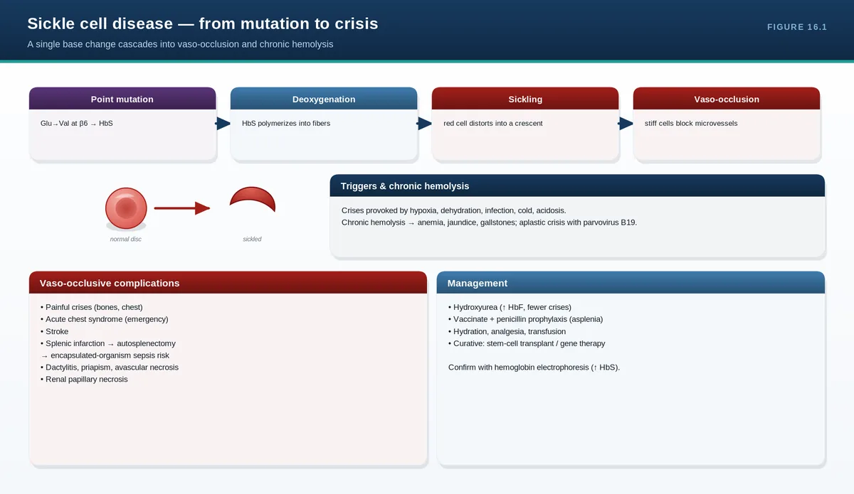
One letter change in the DNA—producing HbS—has enormous consequences. When deoxygenated, HbS polymerizes, deforming red cells into rigid sickles that block small vessels and are destroyed early. Everything clinical flows from these two processes: vaso-occlusion (pain and organ damage) and chronic hemolysis.
Genetics and pathophysiology
Sickle cell disease arises from a single point mutation in the beta-globin gene (glutamate to valine at position 6). Homozygotes (HbSS) have the most severe disease; heterozygotes (HbAS, sickle trait) are generally asymptomatic carriers; compound heterozygotes include HbSC disease and sickle-beta-thalassemia. When oxygen is low, HbS molecules polymerize into rigid fibers that distort the cell; repeated sickling stiffens membranes, promotes adhesion to the endothelium, and triggers vaso-occlusion and inflammation, while the damaged cells undergo hemolysis. Dehydration, acidosis, infection, cold, and hypoxia precipitate sickling.
Acute complications
Vaso-occlusive (pain) crisis: the hallmark—severe bone and tissue pain from microvascular occlusion.
Acute chest syndrome: fever, chest pain, hypoxia, and a new pulmonary infiltrate—a leading cause of death; treat aggressively.
Stroke: especially in children; transcranial Doppler screening identifies risk.
Splenic sequestration: sudden splenic pooling of blood with a falling hemoglobin—life-threatening in young children.
Aplastic crisis: parvovirus B19 shuts down red cell production atop chronic hemolysis.
Infection: functional asplenia raises risk from encapsulated organisms (pneumococcus); a major cause of childhood death.
Priapism: a urological emergency.
Chronic organ damage
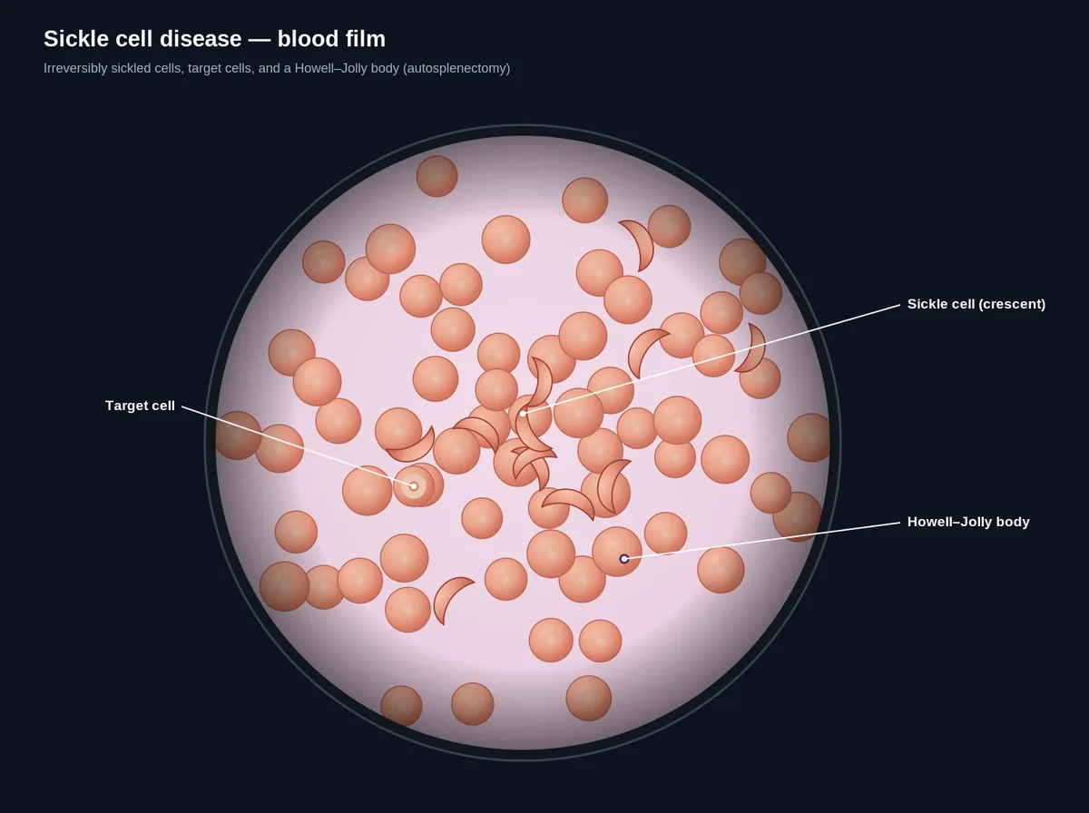
Years of vaso-occlusion and hemolysis damage many organs: the kidney (impaired concentrating ability, papillary necrosis), the retina (proliferative retinopathy), bones (avascular necrosis of the femoral head), the lungs (pulmonary hypertension), and skin (leg ulcers). The spleen progressively infarcts, producing autosplenectomy and Howell-Jolly bodies on the film.
Diagnosis and management
Diagnosis is by newborn screening and hemoglobin electrophoresis/HPLC (HbS predominant with little or no HbA in disease; see Chapter 6), supported by sickle solubility testing. Management combines prevention and treatment: hydroxyurea raises protective HbF and reduces crises and acute chest syndrome; transfusion (simple or exchange) treats and prevents severe complications such as stroke; penicillin prophylaxis and vaccination guard against infection; and acute crises need analgesia, hydration, oxygen, and treatment of triggers. Folate is given for chronic hemolysis. Newer agents (voxelotor, crizanlizumab, L-glutamine) add options, and hematopoietic stem cell transplantation or gene therapy can be curative in selected patients.
PITFALLS & PEARLS
Acute chest syndrome (fever, hypoxia, new infiltrate) is a leading cause of death—recognize and treat it aggressively.
Fever in sickle cell disease is an emergency: functional asplenia means overwhelming pneumococcal sepsis is a real risk.
Hydroxyurea works by raising HbF—it reduces pain crises, acute chest syndrome, and transfusion need.
In acute stroke or severe acute chest syndrome, exchange transfusion (lowering HbS%) is preferred over simple transfusion.
Parvovirus B19 causes an aplastic crisis; sudden splenic enlargement with falling Hb is splenic sequestration—both are emergencies.
CASE VIGNETTE
A child with known HbSS develops fever, chest pain, hypoxia, and a new lobar infiltrate on chest X-ray.
Reasoning: this is acute chest syndrome—a leading cause of mortality. Management includes oxygen, antibiotics, analgesia, and, for severe disease, exchange transfusion to reduce the HbS fraction.
QUICK REVIEW
Single mutation (β6 Glu→Val) → HbS polymerizes when deoxygenated → vaso-occlusion + hemolysis.
Genotypes: SS (severe), AS (trait, asymptomatic), SC and S-β-thal (compound).
Acute: pain crisis, acute chest syndrome, stroke, splenic sequestration, aplastic crisis (parvovirus), infection, priapism.
Chronic: renal, retinal, avascular necrosis, pulmonary hypertension, autosplenectomy.
Manage: hydroxyurea (↑HbF), transfusion/exchange, penicillin + vaccines, analgesia/hydration; HSCT/gene therapy can cure.
Sickle cell diseaseSickle cell disease — blood film
17Acquired Hemolytic Anemias: Autoimmune and Microangiopathictop ↑
THE BIG IDEA
Here the red cell is normal but the environment turns hostile—antibodies coat it (autoimmune hemolysis) or mechanical forces shred it (microangiopathy). The direct antiglobulin test splits the immune from the non-immune; the blood film—spherocytes versus schistocytes—tells the same story visually.
Autoimmune hemolytic anemia (AIHA)
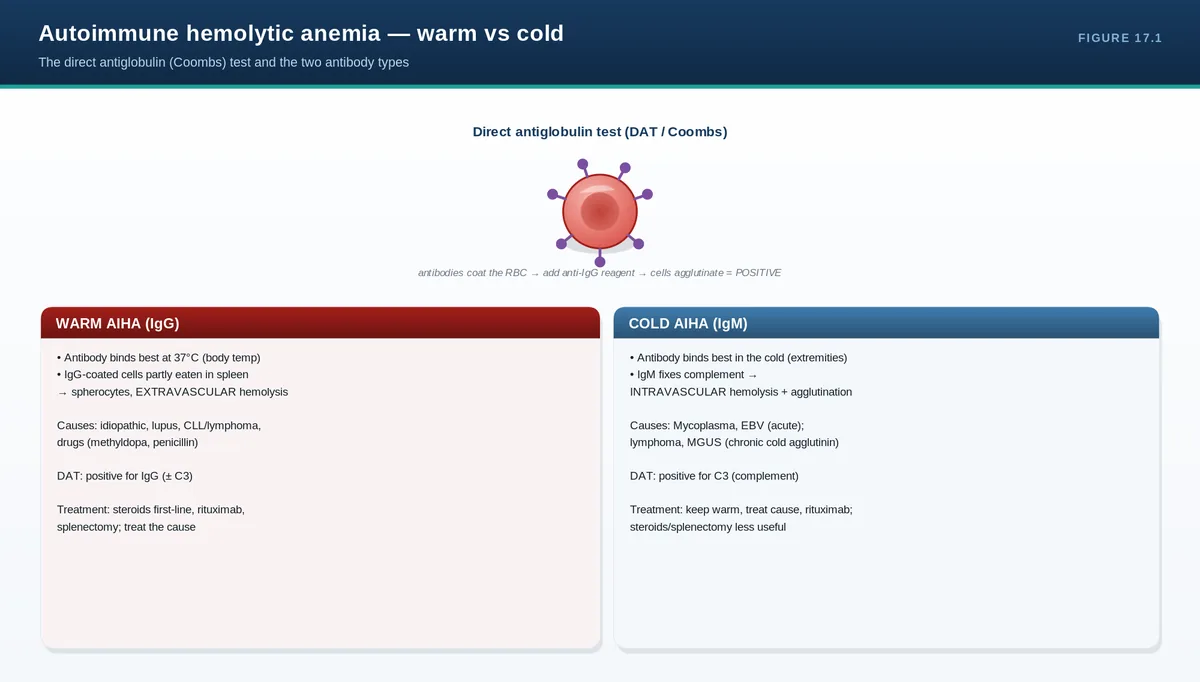
Antibodies against red cell antigens drive destruction, and the temperature at which they bind defines the two types.
Feature
Warm AIHA
Cold AIHA (cold agglutinin)
Antibody
IgG
IgM
Optimal temperature
37°C
Cold (extremities)
DAT pattern
IgG (± C3)
C3 (complement)
Destruction
Extravascular (spleen)
Intravascular / hepatic; agglutination
Film
Spherocytes
Red cell clumping
Associations
Idiopathic, CLL/lymphoma, lupus, drugs
Mycoplasma, EBV, lymphoma
First-line treatment
Corticosteroids
Keep warm; rituximab; treat cause
Warm AIHA is the more common form: IgG-coated cells are removed in the spleen, the film shows spherocytes, and the direct antiglobulin test is positive for IgG. First-line treatment is corticosteroids, with rituximab and splenectomy as later options. Cold agglutinin disease is mediated by IgM that fixes complement in cooler peripheries, causing agglutination and often intravascular hemolysis; steroids work poorly, so management centers on keeping the patient warm, treating any underlying infection or lymphoma, and using rituximab.
Microangiopathic hemolytic anemia (MAHA)
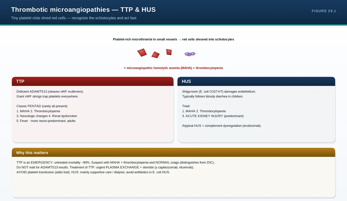
When red cells are forced through vessels partly occluded by microthrombi or damaged endothelium, they fragment into schistocytes. MAHA with thrombocytopenia defines the thrombotic microangiopathies—thrombotic thrombocytopenic purpura (TTP), hemolytic uremic syndrome (HUS), and disseminated intravascular coagulation (DIC)—covered in the thrombosis section. TTP (from severe ADAMTS13 deficiency) is a particular emergency: schistocytes plus thrombocytopenia should trigger urgent plasma exchange, not platelet transfusion. Mechanical heart valves can cause a similar fragmentation hemolysis.
Other acquired causes
Paroxysmal nocturnal hemoglobinuria is an acquired stem cell mutation (PIGA) that strips complement-protective proteins from blood cells, causing intravascular hemolysis, thrombosis, and cytopenias; it is diagnosed by flow cytometry. Infections (notably malaria) and certain drugs are further acquired causes.
PITFALLS & PEARLS
Warm AIHA: DAT positive for IgG, spherocytes, extravascular—treat with steroids. Cold: DAT positive for C3, IgM, keep warm.
Steroids are far less effective in cold agglutinin disease—warmth and treating the underlying cause matter more.
Schistocytes + thrombocytopenia = thrombotic microangiopathy until proven otherwise; consider TTP and act urgently.
In suspected TTP do NOT transfuse platelets reflexively—start plasma exchange.
Consider PNH (flow cytometry) in unexplained intravascular hemolysis with thrombosis or cytopenias.
CASE VIGNETTE
A woman with known CLL develops worsening anemia and jaundice. The film shows spherocytes and the direct antiglobulin test is positive for IgG.
Reasoning: warm autoimmune hemolytic anemia, here secondary to CLL. Treat with corticosteroids and address the underlying lymphoproliferative disorder.
QUICK REVIEW
Acquired hemolysis: immune (warm/cold AIHA) or mechanical (MAHA); DAT separates them.
Warm: IgG, spherocytes, extravascular, steroids. Cold: IgM, complement, agglutination, keep warm/rituximab.
MAHA = schistocytes; with low platelets think TTP/HUS/DIC (emergency).
TTP: schistocytes + thrombocytopenia → urgent plasma exchange, not platelets.
PNH: acquired complement sensitivity → intravascular hemolysis, thrombosis; diagnose by flow cytometry.
The anemias covered here account for the great majority of red cell disease you will encounter. Part III turns to the non-malignant white cell disorders—neutropenia, neutrophil dysfunction, and the reactive leukocytoses and eosinophilia—where the central question shifts from red cell size to whether a count change is reactive or clonal.
Part III covers the non-malignant disorders of white cells—chiefly the neutrophils. Two themes run through it. First, too few functioning neutrophils (whether from low numbers or poor function) means infection risk, and the absolute neutrophil count is the number that matters. Second, when white cell counts are high, the single most useful question is whether the rise is reactive (a normal, polyclonal response to something) or clonal (a primary blood malignancy). The malignant white cell disorders themselves—leukemias, lymphomas, myeloma, and the myeloproliferative and myelodysplastic neoplasms—belong to the companion malignant hematology guide.
18Neutropenia and Neutrophil Function Disorderstop ↑
THE BIG IDEA
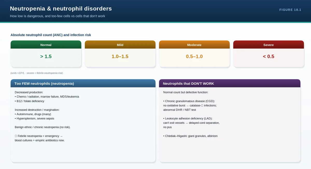
Neutrophils are the front line against bacteria and fungi. When they are too few (neutropenia) or present but dysfunctional, the result is the same: recurrent or severe infection. The number that drives risk is the absolute neutrophil count (ANC), and risk climbs steeply once it falls below 0.5 ×10⁹/L.
The neutrophil and the ANC
Neutrophils mature in the marrow over about a week, then circulate only briefly (hours) before entering tissues. In the blood they exist in two pools—circulating and marginated (stuck to vessel walls)—which is why stress, exercise, or adrenaline can transiently raise the count by demargination. What counts clinically is the absolute neutrophil count: the total WBC multiplied by the percentage of neutrophils (and bands).
Severity of neutropenia
ANC (×10⁹/L)
Infection risk
Mild
1.0–1.5
Minimal
Moderate
0.5–1.0
Moderate
Severe
<0.5
High—urgent
Note benign ethnic neutropenia (associated with the Duffy-null genotype) is very common in Middle East: a mild, lifelong neutropenia with no increased infection risk that should not be over-investigated or mistaken for disease.
Causes of neutropenia
Decreased production: marrow failure or infiltration, chemotherapy and many drugs, B12/folate deficiency, and congenital syndromes (severe congenital neutropenia, cyclic neutropenia).
Increased destruction or margination: immune/autoimmune neutropenia, Felty syndrome, and many infections (especially viral and overwhelming sepsis).
Sequestration: hypersplenism.
Drugs are the single most important acquired cause—chemotherapy predictably, but also idiosyncratic reactions to antithyroid drugs, sulfonamides, antipsychotics (clozapine), and others. A careful medication history is essential.
Clinical features and risk
Neutropenic patients lose the ability to mount pus and localize infection, so signs can be subtle. Mouth ulcers, gingivitis, and perianal infection are common, and bacteremia can progress rapidly. Febrile neutropenia—fever with an ANC below 0.5 ×10⁹/L—is a medical emergency requiring immediate broad-spectrum antibiotics before results return, because untreated sepsis in this setting can be fatal within hours.
Approach and management
Assess with a drug and infection history, examination of mucosa and skin, the trend and chronicity of the count, and a blood film; a bone marrow examination is reserved for unexplained or severe cases. Management is cause-directed: stop the offending drug, treat infection promptly, and use granulocyte colony-stimulating factor (G-CSF) to shorten severe neutropenia or in congenital disease. The cornerstone remains rapid empirical antibiotics for febrile neutropenia.
Qualitative (functional) neutrophil disorders
Here the count is normal but the cells do not work, producing recurrent infections despite adequate numbers. Chronic granulomatous disease results from a defective NADPH oxidase, so neutrophils cannot generate the oxidative burst—patients suffer infections with catalase-positive organisms (Staphylococcus aureus, Aspergillus) and form granulomas; the dihydrorhodamine (or older nitroblue tetrazolium) test is diagnostic. Leukocyte adhesion deficiency prevents neutrophils from leaving the bloodstream, causing delayed umbilical-cord separation, infections without pus, and a high blood neutrophil count. Chediak-Higashi syndrome shows giant granules, partial albinism, and recurrent infection.
PITFALLS & PEARLS
Always calculate the ANC (WBC × % neutrophils)—the percentage alone is misleading.
Febrile neutropenia (fever + ANC <0.5) is an emergency: give broad-spectrum antibiotics immediately, before cultures return.
Take a meticulous drug history—idiosyncratic drug-induced neutropenia (e.g., clozapine, antithyroid drugs) is common and reversible.
Benign ethnic neutropenia (Duffy-null) is mild, lifelong, and harmless—do not over-investigate.
Normal count but recurrent infections (no pus, catalase-positive organisms) points to a FUNCTIONAL defect like chronic granulomatous disease.
CASE VIGNETTE
Ten days after chemotherapy a patient spikes a fever of 38.5°C. The ANC is 0.2 ×10⁹/L. There is no obvious source.
Reasoning: this is febrile neutropenia. Draw cultures but do not wait—start broad-spectrum antibiotics immediately. Delay can be fatal; consider G-CSF and source control as needed.
QUICK REVIEW
Risk tracks the ANC: severe neutropenia is <0.5 ×10⁹/L.
Causes: reduced production (drugs/chemo, marrow, B12/folate, congenital), destruction (immune, infection), sequestration.
Febrile neutropenia = emergency → immediate broad-spectrum antibiotics.
Manage: stop offending drug, treat infection, G-CSF when indicated.
Functional defects (normal count): chronic granulomatous disease, leukocyte adhesion deficiency, Chediak-Higashi.
A high white cell count poses one question above all others: is the rise reactive (a normal, polyclonal response to infection, inflammation, or stress) or clonal (a primary blood malignancy)? The vast majority of leukocytosis is reactive. The clinical context, the pattern of which cell is raised, and the blood film point you toward the answer—and tell you when to escalate.
Definitions and the central question
Leukocytosis is an increased total white cell count; more useful is to name which lineage is raised, because each has its own differential. Reactive (secondary) leukocytosis is a polyclonal response to a stimulus; clonal (primary) leukocytosis comes from a malignant clone.
Neutrophilia is most often reactive—infection, inflammation, corticosteroids, smoking, and physical stress; a left shift and toxic granulation support a reactive cause. Persistent, unexplained neutrophilia warrants screening for a clonal process. Lymphocytosis in children and young adults is usually viral (classically EBV with atypical lymphocytes); in older adults a persistent mature lymphocytosis suggests chronic lymphocytic leukemia, identified by smudge cells and confirmed by flow cytometry. Monocytosis accompanies chronic infections, autoimmune disease, and marrow recovery, but a persistent unexplained rise can signal chronic myelomonocytic leukemia.
Eosinophilia
Eosinophilia is graded mild (0.5–1.5), moderate, and severe; hypereosinophilia is a count above 1.5 ×10⁹/L on repeated testing or in tissue. The causes are best remembered by category: allergy and atopy (asthma, eczema), drug reactions, parasitic and some other infections, connective tissue disease and vasculitis (e.g., eosinophilic granulomatosis with polyangiitis), malignancy (reactive to lymphoma/solid tumors, or clonal in myeloid neoplasms), and adrenal insufficiency. In broad terms, eosinophilia is secondary (reactive, the commonest), primary (clonal, part of a myeloid neoplasm), or idiopathic (hypereosinophilic syndrome).
What makes eosinophilia dangerous is end-organ damage: persistently high eosinophils infiltrate and injure the heart (endomyocardial fibrosis), lungs, skin, and nerves, independent of the cause. The workup confirms persistence, excludes secondary causes (history, parasites, drugs), and then evaluates for a clonal myeloid disorder. Treatment targets the cause; corticosteroids are used for symptomatic or organ-threatening disease—but exclude (or empirically treat) Strongyloides first, because steroids can precipitate fatal hyperinfection.
PITFALLS & PEARLS
For any high count, ask first: reactive or clonal? Most leukocytosis is reactive.
Extreme counts (>100 ×10⁹/L) in acute leukemia → leukostasis, an emergency.
Basophilia is a red flag for a myeloproliferative neoplasm/CML—don't ignore it.
Leukemoid reaction vs CML: use the clinical context, basophilia, and BCR-ABL cytogenetics.
Before giving steroids for eosinophilia, exclude or empirically treat Strongyloides to avoid fatal hyperinfection.
Eosinophil-mediated organ damage (especially cardiac) drives urgency regardless of the underlying cause.
CASE VIGNETTE
A previously healthy 43-year-old woman has a WBC of 12 ×10⁹/L with a normal differential, unchanged on repeat three weeks later; she is asymptomatic with normal hematocrit and platelets.
Reasoning: a mild, stable, asymptomatic leukocytosis with a normal differential and normal other lines is almost always reactive (here, likely smoking or low-grade stress). It needs context and follow-up, not an aggressive malignancy workup.
QUICK REVIEW
Leukocytosis: name the lineage, then ask reactive vs clonal.
Neutrophilia: usually reactive (infection, steroids, smoking); persistent unexplained → screen clonal.
Lymphocytosis: viral in young; CLL (smudge cells, flow) in older adults. Basophilia → think MPN/CML.
Eosinophilia categories: allergy, drugs, parasites, connective tissue/vasculitis, malignancy, adrenal insufficiency.
Organ damage (heart, lung) drives treatment; exclude Strongyloides before steroids.
Part IV turns from cells to clotting. Hemostasis is a staged system—platelets and vessels stop bleeding quickly (primary hemostasis), the coagulation cascade lays down a stable fibrin clot (secondary hemostasis), and fibrinolysis later clears it. Every bleeding disorder is a defect in one of these stages, and the pattern of bleeding usually tells you which. These chapters build that framework and then work through the disorders of platelets and clotting factors. Thrombosis and anticoagulation follow in Part V.
Hemostasis is a controlled, staged response to vascular injury. Platelets and the vessel wall plug the leak within seconds (primary hemostasis); the coagulation cascade then reinforces the plug with a mesh of fibrin (secondary hemostasis); and once healing is underway, fibrinolysis dissolves the clot. Learn the three stages and every bleeding—and clotting—disorder finds its place.
Primary hemostasis
When a vessel is injured it first constricts. Platelets then adhere to exposed subendothelial collagen, using von Willebrand factor (VWF) as the molecular bridge between collagen and the platelet GpIb receptor. Adhesion activates the platelets: they change shape, release the contents of their granules (recruiting more platelets), and link to one another through GpIIb/IIIa receptors that bind fibrinogen. The result is a platelet plug. Defects at this stage—low platelet numbers, poor platelet function, or VWF deficiency—cause mucocutaneous bleeding.
Secondary hemostasis
In parallel, the coagulation cascade generates thrombin, which converts fibrinogen into fibrin to stabilize the plug. Traditionally the cascade is drawn as two arms feeding a common pathway, and this still maps neatly onto the screening tests: the intrinsic pathway (factors XII, XI, IX, VIII) is measured by the aPTT, the extrinsic pathway (factor VII with tissue factor) by the PT, and both converge on the common pathway (factors X, V, II/prothrombin, and fibrinogen). Physiologically, coagulation is better understood as a cell-based process: tissue factor initiates a small amount of thrombin, which amplifies the response on the activated platelet surface and drives a thrombin burst that produces a robust fibrin clot. Factors II, VII, IX, and X (and the regulators protein C and S) depend on vitamin K.
Screening test
Pathway
Factors assessed
Prolonged by
PT (INR)
Extrinsic + common
VII, X, V, II, fibrinogen
Warfarin, liver disease, vitamin K deficiency
aPTT
Intrinsic + common
XII, XI, IX, VIII, X, V, II, fibrinogen
Heparin, hemophilia, VWD, lupus anticoagulant
Regulation and fibrinolysis
Clotting is kept in check by natural anticoagulants—antithrombin, the protein C/protein S system, and tissue factor pathway inhibitor—which confine the clot to the site of injury (their deficiency causes thrombosis, covered in Part V). Later, plasmin breaks fibrin down into fragments, including D-dimer, restoring normal flow. Bleeding can therefore result not only from too little clotting but occasionally from excessive fibrinolysis.
Mucocutaneous bleeding = primary hemostasis (platelets/VWF); deep, delayed bleeding = secondary hemostasis (coagulation factors).
PT screens the extrinsic/common pathway (VII); aPTT screens the intrinsic/common pathway (VIII, IX).
Vitamin K–dependent factors are II, VII, IX, X (and proteins C and S)—the basis of warfarin's action.
Petechiae are a platelet sign, not a coagulation-factor sign—hemophiliacs do not get petechiae.
CASE VIGNETTE
Two students compare bleeding histories: one has lifelong petechiae and nosebleeds; the other has recurrent painful knee swellings after minor knocks.
Reasoning: the first describes a primary-hemostasis (platelet/VWF) pattern; the second describes deep bleeding typical of a coagulation-factor problem such as hemophilia. The pattern alone narrows the diagnosis before any test.
QUICK REVIEW
Three stages: primary (platelet plug), secondary (fibrin via cascade), fibrinolysis (plasmin).
Primary: VWF bridges platelet GpIb to collagen; GpIIb/IIIa binds fibrinogen for aggregation.
Secondary: PT = extrinsic (VII); aPTT = intrinsic (VIII, IX); common pathway X, V, II, fibrinogen.
Vitamin K factors: II, VII, IX, X (+ protein C/S).
Bleeding pattern localizes the defect: mucocutaneous (primary) vs deep/delayed (secondary).
The bleeding history is the single most powerful diagnostic tool—more than any screening test. Decide whether the pattern is mucocutaneous or deep, whether it is lifelong (inherited) or new (acquired), and how severe it is; then a short panel of tests confirms where in hemostasis the problem lies.
History: what to ask
Ask where bleeding occurs (skin and mucosa versus joints and muscles), how long it has been a problem (since childhood suggests an inherited disorder; recent onset suggests an acquired one), and how the patient handled hemostatic challenges—dental extractions, surgery, childbirth, circumcision, and menstruation. A lifelong history of bleeding only with major challenges differs from spontaneous bleeding. Family history points to inherited disorders and their inheritance pattern, and a drug history is essential: antiplatelet agents, anticoagulants, and many over-the-counter drugs cause or unmask bleeding. Structured bleeding-assessment scores help quantify whether the history is truly abnormal.
Recognizing the pattern
Clue
Suggests primary hemostasis
Suggests secondary hemostasis
Bleeding sites
Skin, mucous membranes
Joints, muscles, deep tissue
Petechiae
Present
Absent
Timing
Immediate after injury
Delayed; rebleeding
Typical causes
Thrombocytopenia, platelet dysfunction, VWD
Hemophilia, factor deficiency, anticoagulants
Testing strategy
Start with the screen: a platelet count, PT, aPTT, and fibrinogen, alongside a blood film. Interpret these with the master algorithm from Chapter 6—if a clotting time is prolonged, a mixing study separates factor deficiency (corrects) from an inhibitor (does not). Then move to specific tests: von Willebrand studies, individual factor assays, and platelet function testing. A crucial point: a normal screen does not exclude a bleeding disorder. Von Willebrand disease, mild factor deficiencies, platelet function defects, and factor XIII deficiency can all bleed with a completely normal platelet count, PT, and aPTT—so a convincing history justifies specialized testing even when the screen is normal.
PITFALLS & PEARLS
A normal PT, aPTT, and platelet count do NOT exclude a bleeding disorder—VWD, mild factor deficiency, platelet dysfunction, and factor XIII deficiency are all missed by the screen.
Lifelong bleeding since childhood points to an inherited disorder; new bleeding in an older adult suggests an acquired cause (drugs, acquired inhibitor, liver disease).
Always take a full drug history—antiplatelets and anticoagulants are the commonest cause of bleeding.
Use the mixing study to split a prolonged clotting time into deficiency (corrects) versus inhibitor (does not).
CASE VIGNETTE
A 19-year-old woman has had heavy periods, easy bruising, and prolonged bleeding after wisdom-tooth removal since adolescence. Platelet count, PT, and aPTT are all normal.
Reasoning: a lifelong mucocutaneous bleeding history with a normal screen is classic for von Willebrand disease. The normal screen must not reassure—order VWF studies.
QUICK REVIEW
History is the most powerful test: pattern (mucocutaneous vs deep), onset (lifelong vs new), severity, drugs, family.
Screen: platelet count, PT, aPTT, fibrinogen, film; mixing study if a time is prolonged.
Then specific tests: VWF studies, factor assays, platelet function testing.
A normal screen does NOT exclude VWD, mild factor deficiency, platelet dysfunction, or factor XIII deficiency.
Inherited (lifelong) vs acquired (new) shapes the differential.
Approach to the bleeding patient
22Thrombocytopenia and Immune Thrombocytopenia (ITP)top ↑
THE BIG IDEA
A low platelet count comes from one of three mechanisms—reduced production, increased destruction, or sequestration. The first move is always to look at the blood film: confirm the count is real (not clumping), and exclude the dangerous mimics (TTP, DIC, HIT). Immune thrombocytopenia (ITP) is then a diagnosis of exclusion in an otherwise well patient.
Two artifacts and three emergencies must be addressed up front. The artifact is pseudothrombocytopenia from EDTA-induced platelet clumping—always confirm a low count on the film. The emergencies are thrombotic thrombocytopenic purpura (schistocytes), disseminated intravascular coagulation (abnormal clotting times, low fibrinogen), and heparin-induced thrombocytopenia (recent heparin, thrombosis)—all covered in Part V and all needing specific, urgent action.
Immune thrombocytopenia (ITP)
ITP is caused by autoantibodies that both accelerate platelet destruction and impair their production. It may be primary or secondary to another condition (HIV, hepatitis C, lupus, chronic lymphocytic leukemia, certain drugs, and H. pylori). The presentation differs by age: in children it is often acute and post-viral and resolves spontaneously, whereas in adults it tends to be chronic. The hallmark is isolated thrombocytopenia—a low platelet count with otherwise normal blood lines and film—in a patient who is clinically well apart from mucocutaneous bleeding. There is no confirmatory test; ITP remains a diagnosis of exclusion.
Management
Treat the patient, not just the number—many patients with moderate thrombocytopenia and no bleeding can be observed. Treatment is indicated for significant bleeding or very low counts. First-line therapy is corticosteroids, with intravenous immunoglobulin (IVIG) added when a faster rise is needed (for example before a procedure or with active bleeding). Second-line options include thrombopoietin-receptor agonists, rituximab, and splenectomy. Life-threatening bleeding is treated with combined platelet transfusion, IVIG, and corticosteroids.
PITFALLS & PEARLS
Confirm every unexpected low platelet count on the film first—pseudothrombocytopenia (clumping) is common and harmless.
Exclude TTP, DIC, and HIT before settling on ITP—they are emergencies with specific treatments.
ITP is isolated thrombocytopenia in a well patient and remains a diagnosis of exclusion.
Treat based on bleeding and count together, not the number alone—asymptomatic moderate ITP is often just observed.
Avoid routine platelet transfusion in ITP unless there is severe bleeding—transfused platelets are rapidly destroyed.
CASE VIGNETTE
A well 28-year-old woman has scattered petechiae and a platelet count of 12 ×10⁹/L; hemoglobin, white count, and the film are otherwise normal with no schistocytes.
Reasoning: isolated severe thrombocytopenia in an otherwise well patient, with a normal film, is ITP once mimics are excluded. First-line treatment is corticosteroids, adding IVIG if rapid correction is needed.
QUICK REVIEW
Low platelets: decreased production, increased destruction, or sequestration—check the film first.
Exclude pseudothrombocytopenia (clumping) and the emergencies TTP, DIC, HIT.
ITP = isolated thrombocytopenia, well patient, diagnosis of exclusion; children acute/self-limited, adults chronic.
Treat for bleeding or very low counts: steroids ± IVIG first line; TPO agonists, rituximab, splenectomy later.
Don't reflexively transfuse platelets in ITP—reserve for severe bleeding.
Sometimes the platelets are present in normal numbers but simply do not work. The result is mucocutaneous bleeding with a normal platelet count and normal clotting times—a picture that is easy to miss if you only look at numbers. Acquired causes, above all drugs like aspirin, are far more common than the inherited disorders.
How platelets can fail
Recall the steps of platelet function: adhesion to the vessel wall (via GpIb and VWF), activation and shape change, secretion of granule contents, and aggregation (via GpIIb/IIIa and fibrinogen). A defect at any step impairs the platelet plug. Because the platelet count and the coagulation screen are normal, these disorders are diagnosed by specialized platelet function testing.
Acquired platelet dysfunction (common)
Drugs are the leading cause. Aspirin irreversibly inhibits cyclooxygenase, blunting platelet aggregation for the life of the platelet; P2Y12 inhibitors (clopidogrel) and other antiplatelet agents act on different steps, and NSAIDs cause reversible inhibition. Systemic conditions also impair platelets: uremia (kidney failure), liver disease, cardiopulmonary bypass, and myeloproliferative neoplasms. A careful drug and comorbidity history usually identifies the cause.
Inherited platelet disorders (rare but instructive)
Disorder
Defect
Clue
Bernard-Soulier syndrome
GpIb (adhesion to VWF)
Large platelets + mild thrombocytopenia
Glanzmann thrombasthenia
GpIIb/IIIa (aggregation)
Normal count and platelet size; failed aggregation, severe bleeding
Storage pool / granule defects
Deficient granule content (e.g., gray platelet)
Mild bleeding; abnormal secretion
Diagnosis and management
Diagnosis rests on platelet function testing—light transmission aggregometry is the reference method, showing characteristic patterns (for example, absent aggregation to all agonists in Glanzmann thrombasthenia). The coagulation screen is normal. Management starts with removing the offending drug and treating any underlying condition; bleeding is reduced with desmopressin (which improves platelet function in some disorders), antifibrinolytic agents (tranexamic acid), and—for severe bleeding or surgery—platelet transfusion.
PITFALLS & PEARLS
Mucocutaneous bleeding with a NORMAL platelet count and NORMAL clotting times points to a platelet function defect (or VWD).
Aspirin's effect is irreversible and lasts the platelet's lifespan (~7–10 days)—stop it well before surgery.
Uremia is a common, reversible cause of platelet dysfunction—dialysis and desmopressin help.
Bernard-Soulier has big platelets and a low-normal count; Glanzmann has a normal count—both bleed despite the numbers.
Tranexamic acid is a useful, simple adjunct for mucosal bleeding and dental procedures.
CASE VIGNETTE
A patient on long-term aspirin and an NSAID for arthritis has easy bruising and prolonged bleeding after a dental extraction; platelet count, PT, and aPTT are all normal.
Reasoning: acquired platelet dysfunction from antiplatelet/NSAID therapy. The screen is normal because the problem is platelet function, not number or the cascade. Manage by stopping the drugs where possible and using tranexamic acid for the procedure.
QUICK REVIEW
Normal count, normal screen, mucocutaneous bleeding → platelet function defect (or VWD).
Acquired (common): aspirin (irreversible COX), P2Y12 inhibitors, NSAIDs, uremia, liver disease, bypass.
Inherited (rare): Bernard-Soulier (GpIb, large platelets), Glanzmann (GpIIb/IIIa), granule/storage pool defects.
Diagnose by platelet aggregometry; coagulation screen normal.
Manage: stop offending drugs, desmopressin, tranexamic acid, platelets if severe.
Von Willebrand factor (VWF) does two jobs: it glues platelets to the injured vessel wall, and it carries and protects factor VIII in the circulation. When VWF is deficient or dysfunctional, both jobs suffer—producing mucocutaneous bleeding and sometimes a low factor VIII. It is the most common inherited bleeding disorder.
VWF biology
VWF is made by endothelial cells and megakaryocytes and circulates as large multimers—the bigger the multimer, the more effective. It performs primary hemostasis by bridging the platelet GpIb receptor to subendothelial collagen, and it acts as a chaperone for factor VIII, prolonging its half-life. This dual role explains the laboratory picture: severe VWF deficiency lowers factor VIII enough to prolong the aPTT, even though the primary problem is in primary hemostasis.
Types
Type
Defect
Notes
Type 1
Partial quantitative deficiency
Most common (~75%); usually mild
Type 2
Qualitative (dysfunctional VWF)
Subtypes 2A, 2B, 2M, 2N
Type 3
Near-complete deficiency
Severe; very low factor VIII
Two subtypes are worth remembering. Type 2B has a gain-of-function VWF that binds platelets in the circulation, causing thrombocytopenia. Type 2N has VWF that cannot bind factor VIII, so factor VIII is low and the picture mimics hemophilia A (but with autosomal, not X-linked, inheritance).
Clinical features
Inheritance is usually autosomal, so unlike hemophilia it affects both sexes. Bleeding is mucocutaneous: epistaxis, easy bruising, heavy menstrual bleeding, and bleeding after dental work or surgery. Severity varies widely, and mild disease may only show up under hemostatic challenge.
Diagnosis and management
Testing measures VWF antigen (how much VWF), VWF activity (how well it works—ristocetin cofactor or GpIb-binding assays), and factor VIII; multimer analysis defines the subtype. The platelet count is usually normal (except type 2B), and the aPTT may be normal or prolonged depending on the factor VIII level. Management depends on type and severity: desmopressin releases stored VWF and is effective in many type 1 patients (test responsiveness first; avoid in type 2B, where it can worsen thrombocytopenia), while VWF-containing concentrates are used for severe disease, type 3, and surgery. Antifibrinolytics help with mucosal bleeding, and pregnancy and procedures need specific planning.
PITFALLS & PEARLS
VWD is the most common inherited bleeding disorder and is autosomal—it affects men and women alike.
Severe VWD lowers factor VIII (VWF is its chaperone), so the aPTT can be prolonged even though the core defect is in primary hemostasis.
Type 2B causes thrombocytopenia and is the exception where desmopressin is contraindicated.
Type 2N mimics hemophilia A (low factor VIII) but is inherited autosomally—test VWF before labeling a patient hemophiliac.
Desmopressin works in many type 1 patients—confirm a response with a trial before relying on it for surgery.
CASE VIGNETTE
A 23-year-old woman has lifelong heavy periods, frequent nosebleeds, and a mother with similar symptoms. VWF antigen and activity are both reduced; factor VIII is low-normal; platelets are normal.
Reasoning: an autosomal, lifelong mucocutaneous bleeding history with low VWF antigen and activity is type 1 von Willebrand disease. Desmopressin (after a trial) and tranexamic acid manage menstrual and procedural bleeding.
QUICK REVIEW
VWF bridges platelets to collagen (primary hemostasis) and carries factor VIII.
Most common inherited bleeding disorder; autosomal (both sexes); mucocutaneous bleeding.
Types: 1 (partial quantitative, mild), 2 (qualitative: 2A/2B/2M/2N), 3 (severe, very low FVIII).
Tests: VWF antigen, VWF activity (ristocetin cofactor), factor VIII, multimers; platelets usually normal (not 2B).
Treat: desmopressin (not in 2B), VWF concentrates, tranexamic acid.
Von Willebrand disease
25Hemophilia, Factor Deficiencies, and Rare Bleeding Disorderstop ↑
THE BIG IDEA
Hemophilia A (factor VIII deficiency) and hemophilia B (factor IX deficiency) are X-linked disorders that cause deep bleeding—into joints and muscles—with an isolated prolonged aPTT. Factor replacement (and now non-factor therapies) prevents the crippling joint disease that defined the condition for centuries. The major modern complication is the development of inhibitors.
Hemophilia A and B
Both are inherited in an X-linked recessive pattern, so they predominantly affect males, with females usually carriers (who can nonetheless bleed if factor levels are low). Hemophilia A (factor VIII) is far more common than hemophilia B (factor IX); clinically the two are indistinguishable. Severity tracks the residual factor level.
Severity
Factor level
Bleeding pattern
Severe
<1%
Spontaneous hemarthrosis and muscle bleeds
Moderate
1–5%
Bleeding with minor trauma
Mild
5–40%
Bleeding mainly with surgery or major trauma
The hallmark is deep bleeding: recurrent hemarthrosis (often into the same target joints, leading to chronic arthropathy), muscle hematomas, and, dangerously, intracranial and retroperitoneal bleeds; surgical and dental bleeding is delayed and prolonged. The laboratory shows a prolonged aPTT with a normal PT, platelet count, and VWF; the aPTT corrects on a mixing study (a deficiency, not an inhibitor), and a specific factor assay distinguishes A from B.
Management
Treatment replaces the missing factor, either on demand for bleeds or as regular prophylaxis to prevent hemarthrosis in severe disease. For hemophilia A, the non-factor agent emicizumab (a bispecific antibody that mimics factor VIII) has transformed prophylaxis, and desmopressin can raise factor VIII enough to cover minor procedures in mild disease. Antifibrinolytics are useful adjuncts for mucosal and dental bleeding, and gene therapy is emerging. The feared complication is the development of inhibitors—alloantibodies against the infused factor that render replacement ineffective and require bypassing agents and immune tolerance strategies.
Acquired hemophilia
Acquired hemophilia is a separate entity: autoantibodies against factor VIII arise in a previously normal person—often an older adult or postpartum woman—causing sudden, severe bleeding. The aPTT is prolonged and, crucially, does NOT correct on mixing (an inhibitor). Treatment combines control of bleeding with bypassing agents and immunosuppression to remove the antibody.
Other factor deficiencies and rare disorders
Factor XI deficiency (common in Ashkenazi Jews) causes variable, often trauma-related bleeding. Factor XIII deficiency is unique: it stabilizes the clot, so the PT and aPTT are normal but patients have delayed bleeding, classically umbilical-stump bleeding in neonates and a risk of intracranial hemorrhage. Fibrinogen disorders, factor VII deficiency, and combined deficiencies also occur. Acquired multifactor problems are far more common in practice: vitamin K deficiency (factors II, VII, IX, X) and liver disease (most factors), both prolonging the PT. Tranexamic acid is a valuable adjunct across many of these bleeding disorders.
PITFALLS & PEARLS
Hemophilia: isolated prolonged aPTT (normal PT, platelets, VWF) that corrects on mixing—deep bleeding, X-linked.
Distinguish A (factor VIII) from B (factor IX) by specific factor assays; clinically identical.
Inhibitors are the major complication of replacement therapy—suspect them when bleeds stop responding to factor.
Acquired hemophilia: new severe bleeding in an older or postpartum patient, aPTT that does NOT correct on mixing.
Factor XIII deficiency has a NORMAL PT and aPTT but causes delayed bleeding (umbilical stump, intracranial)—remember it when the screen is normal.
CASE VIGNETTE
A 2-year-old boy has recurrent painful, swollen knees and a large bruise after minor falls. The aPTT is prolonged, the PT and platelets are normal, and the aPTT corrects on mixing; factor VIII is <1%.
Reasoning: deep joint bleeding in a boy with an isolated, correctable prolonged aPTT and very low factor VIII is severe hemophilia A. Management is factor VIII (or emicizumab) prophylaxis to prevent arthropathy.
QUICK REVIEW
Hemophilia A (FVIII) and B (FIX): X-linked, deep bleeding (hemarthrosis), isolated prolonged aPTT that corrects on mixing.
Severity by factor level: <1% severe, 1–5% moderate, 5–40% mild.
Treat: factor replacement / emicizumab (A), desmopressin for mild A, tranexamic acid; watch for inhibitors.
Acquired hemophilia: FVIII autoantibody, older/postpartum, aPTT does NOT correct on mixing.
Rare: factor XI (Ashkenazi), factor XIII (normal PT/aPTT, delayed/umbilical bleeding); vitamin K deficiency and liver disease prolong PT.
Part IV asked why patients bleed; Part V asks the opposite—why they clot when they should not, and how we safely prevent and treat that. The same hemostatic system, pushed the other way, produces venous and arterial thrombosis. These chapters cover the approach to thrombosis and inherited thrombophilia, the anticoagulant and antiplatelet drugs you will prescribe and reverse, and two disorders where low platelets paradoxically signal clotting: heparin-induced thrombocytopenia and the thrombotic microangiopathies
Thrombosis is clotting in the wrong place. Virchow's triad—stasis, endothelial injury, and a hypercoagulable state—explains why it happens, and almost every clot reflects some combination of the three. Most venous thromboembolism is provoked by an identifiable risk factor, which is why thrombophilia testing rarely changes what you do.
Virchow's triad and the spectrum of thrombosis
Venous thromboembolism (VTE) spans deep vein thrombosis (DVT) and pulmonary embolism (PE)—the same disease at two sites. Venous clots are fibrin-rich red clots that form in slow-flowing blood and are treated with anticoagulants; arterial clots are platelet-rich white clots that form on ruptured plaques and are treated with antiplatelet agents. Knowing which kind of clot you are facing guides the whole approach.
Risk factors: provoked versus unprovoked
The first question in any VTE is whether it was provoked. Provoking factors include recent surgery, immobility or hospitalization, active cancer, pregnancy and the postpartum period, estrogen therapy, trauma, and long-haul travel. A clot with a clear, reversible provoker behaves differently—and is treated for a shorter time—than an unprovoked clot, which carries a higher recurrence risk. Active cancer deserves special mention as both a strong provoker and a reason for extended anticoagulation.
Inherited and acquired thrombophilia
Thrombophilia
Nature
Note
Factor V Leiden
Inherited (most common)
Activated protein C resistance
Prothrombin G20210A
Inherited
Raised prothrombin level
Protein C or S deficiency
Inherited
Loss of natural anticoagulant
Antithrombin deficiency
Inherited
Strong risk; heparin may work poorly
Antiphospholipid syndrome
Acquired
Arterial AND venous clots; pregnancy loss
When to test—and the traps
Resist reflexive thrombophilia screening. For most provoked clots, the result does not change management—the duration of anticoagulation is decided by whether the clot was provoked and by bleeding risk, not by an inherited mutation. Testing also has timing pitfalls: an acute clot and anticoagulant drugs distort many assays (protein C/S and antithrombin levels fall acutely; heparin and warfarin interfere), so tests done at the wrong moment mislead. The important exception is antiphospholipid syndrome, which is acquired, causes both arterial and venous thrombosis and pregnancy loss, and does change management (often indefinite anticoagulation, with warfarin preferred over DOACs).
Diagnosis of VTE
Combine clinical probability with testing. A validated score (such as the Wells score) sets the pretest probability. A D-dimer is sensitive but not specific—useful to rule out VTE when probability is low (a normal D-dimer excludes it) but unhelpful when probability is high. Imaging confirms: compression ultrasound for DVT and CT pulmonary angiography for PE.
PITFALLS & PEARLS
Venous clots (fibrin-rich) → anticoagulants; arterial clots (platelet-rich) → antiplatelets. Match the drug to the clot.
Most VTE is provoked—thrombophilia testing rarely changes management and is often best avoided.
Protein C/S and antithrombin fall during acute clot and with anticoagulants—don't test at the wrong time.
Antiphospholipid syndrome is the thrombophilia that matters most: it causes arterial and venous clots and pregnancy loss and alters treatment.
A normal D-dimer helps rule out VTE only when pretest probability is low; it cannot rule it in.
CASE VIGNETTE
A 30-year-old woman on the combined oral contraceptive develops a swollen, painful calf two weeks after a long flight. Ultrasound confirms a DVT.
Reasoning: this is provoked VTE (estrogen plus immobility—two arms of Virchow's triad). Treat with anticoagulation and address the provoker; routine thrombophilia testing is not needed and would not change management.
QUICK REVIEW
Virchow's triad: stasis, endothelial injury, hypercoagulability. VTE = DVT + PE.
Provoked (surgery, immobility, cancer, estrogen, pregnancy) vs unprovoked (higher recurrence).
Inherited: factor V Leiden (commonest), prothrombin G20210A, protein C/S and antithrombin deficiency.
Antiphospholipid syndrome (acquired): arterial + venous clots, pregnancy loss—does change management.
Diagnose with pretest probability + D-dimer (rules out if low) + imaging (US, CTPA).
Two drug families, two targets. Antiplatelet agents blunt primary hemostasis and are used for arterial (platelet-rich) clots; anticoagulants block the coagulation cascade and are used for venous (fibrin-rich) clots. Choosing well means matching the drug to the clot and the patient—and always knowing how to reverse it.
Antiplatelet agents
Aspirin irreversibly inhibits cyclooxygenase, reducing platelet aggregation for the platelet's lifespan. P2Y12 inhibitors (clopidogrel, ticagrelor, prasugrel) block ADP-mediated activation, and glycoprotein IIb/IIIa inhibitors are potent intravenous agents used in selected acute settings. These drugs are the mainstay for arterial disease—acute coronary syndromes, ischemic stroke, and coronary stents (often as dual antiplatelet therapy).
Anticoagulants
Drug class
Target / mechanism
Monitoring
Reversal
Unfractionated heparin
Antithrombin → IIa & Xa
aPTT (or anti-Xa)
Protamine sulfate
LMWH (enoxaparin)
Antithrombin → mainly Xa
Usually none (anti-Xa if needed)
Partial (protamine)
Warfarin
Vitamin K antagonist (II, VII, IX, X)
INR
Vitamin K, PCC, FFP
DOAC – dabigatran
Direct thrombin (IIa)
None routine
Idarucizumab
DOAC – apixaban/rivaroxaban
Direct factor Xa
None routine
Andexanet alfa; PCC
A few practical points anchor these drugs. Heparins act immediately and are used to initiate treatment; low-molecular-weight heparin has predictable pharmacokinetics and rarely needs monitoring. Warfarin is cheap and reversible but has a narrow therapeutic window, countless food and drug interactions, and a delayed onset—so it requires overlapping (bridging) cover when started for acute clot. Direct oral anticoagulants (DOACs) are now first-line for most VTE and atrial fibrillation: fixed dosing, no routine monitoring, and specific reversal agents.
Choosing therapy and special populations
Treatment duration for VTE depends on the provoker: a provoked clot is typically treated for about three months, while an unprovoked clot or cancer-associated thrombosis often warrants extended therapy. Special situations matter: in pregnancy, low-molecular-weight heparin is used because warfarin is teratogenic and DOACs are avoided; in significant renal impairment, dosing must be adjusted (DOACs and LMWH are renally cleared); and antiphospholipid syndrome favors warfarin over DOACs. The universal complication is bleeding, which is why knowing each agent's reversal is essential.
PITFALLS & PEARLS
Antiplatelets for arterial disease; anticoagulants for venous thromboembolism—don't swap them.
Warfarin has a delayed onset and transient early hypercoagulability—bridge with heparin when treating acute clot.
Protamine reverses heparin (fully) and LMWH (partially); vitamin K/PCC reverse warfarin; idarucizumab reverses dabigatran; andexanet reverses Xa inhibitors.
In pregnancy use LMWH—warfarin is teratogenic and DOACs are not recommended.
DOACs are first-line for most VTE and atrial fibrillation, but avoid them in antiphospholipid syndrome and severe renal failure.
CASE VIGNETTE
A 55-year-old man with a first unprovoked DVT and normal renal function asks about treatment options and how long he will need them.
Reasoning: a DOAC is first-line, with no routine monitoring. Because the clot is unprovoked, extended anticoagulation is considered after weighing recurrence against bleeding risk—not the fixed three months used for provoked clots.
HIT is the great paradox of hematology: an immune reaction to heparin that causes thrombosis even as the platelet count falls. The danger is clotting, not bleeding. Recognizing it, stopping all heparin, and starting a non-heparin anticoagulant—while avoiding the classic traps—can save a limb or a life.
Pathophysiology
Heparin can bind platelet factor 4 (PF4) to form a complex that the immune system attacks. The resulting IgG antibodies bind the PF4-heparin complex and cross-link platelet receptors, activating platelets en masse. This both consumes platelets (the thrombocytopenia) and generates a powerfully prothrombotic state (the thrombosis). It is counterintuitive precisely because the falling platelet count signals clotting, not bleeding.
Clinical recognition: the 4Ts
Suspect HIT using the 4Ts: the Timing (platelet fall typically 5–10 days after starting heparin, or faster on re-exposure), the degree of Thrombocytopenia (usually a fall of more than 50%, with a nadir rarely below 20 ×10⁹/L), new Thrombosis (venous or arterial), and the absence of another (oTher) explanation. Crucially, patients clot rather than bleed; bleeding is unusual, and the thrombosis can be limb- or life-threatening.
Diagnosis
Use the 4Ts score to gauge probability, then test. A PF4 immunoassay (anti-PF4/heparin antibodies) is highly sensitive—a negative result largely excludes HIT—but not specific, so a positive immunoassay in a high-probability patient is confirmed with a functional platelet-activation assay (such as the serotonin release assay), which is specific. Do not wait for confirmation to act if suspicion is high.
Management—and the traps
Stop all heparin immediately: including flushes and LMWH—but note that stopping heparin alone is NOT sufficient treatment.
Start a non-heparin anticoagulant: argatroban, fondaparinux, or a DOAC—because the patient remains intensely prothrombotic.
Do NOT start warfarin until platelets recover: early warfarin can precipitate venous limb gangrene by dropping protein C; overlap and delay until the count normalizes.
Avoid platelet transfusions: they add fuel to a prothrombotic fire and are reserved for serious bleeding only.
Never re-expose: document the diagnosis and avoid all heparin in the future.
PITFALLS & PEARLS
HIT causes THROMBOSIS despite thrombocytopenia—the risk is clotting, not bleeding.
Stopping heparin alone is inadequate—you MUST start a non-heparin anticoagulant.
Do not give warfarin until platelets recover (risk of venous limb gangrene); avoid platelet transfusions.
A negative PF4 immunoassay essentially rules HIT out; confirm a positive one with a functional assay (SRA).
Use the 4Ts score to decide who to test and treat—timing is classically 5–10 days after heparin exposure.
CASE VIGNETTE
On day 7 of unfractionated heparin prophylaxis, a postoperative patient's platelet count falls from 250 to 90 ×10⁹/L and a new DVT is found. There is no bleeding.
Reasoning: timing, a >50% platelet fall, and new thrombosis give a high 4Ts score—HIT. Stop all heparin and start a non-heparin anticoagulant (e.g., argatroban or fondaparinux); do not start warfarin until platelets recover, and do not transfuse platelets.
QUICK REVIEW
HIT: IgG vs PF4-heparin complexes activate platelets → thrombosis with a falling platelet count.
Recognize with the 4Ts: Timing (5–10 days), Thrombocytopenia (>50% fall), Thrombosis, no oTher cause.
Diagnose: 4Ts → PF4 immunoassay (sensitive) → functional SRA (specific).
Treat: stop ALL heparin AND start a non-heparin anticoagulant (argatroban, fondaparinux, DOAC).
Traps: no early warfarin (limb gangrene), no platelet transfusion, never re-expose.
Thrombotic microangiopathies (TMAs) are disorders in which tiny vessels fill with platelet thrombi, shredding red cells as they pass (microangiopathic hemolysis) and consuming platelets, while the resulting ischemia injures organs. The central pattern is the same everywhere—schistocytes plus thrombocytopenia plus organ damage—and the key fork is TTP versus HUS. TTP is an emergency.
Recognizing a TMA
The unifying triad is microangiopathic hemolytic anemia (anemia with schistocytes, high LDH, low haptoglobin), thrombocytopenia, and organ injury—without a coagulopathy. That last point is important: in TTP and HUS the PT, aPTT, and fibrinogen are typically normal, which separates them from disseminated intravascular coagulation. Any patient with schistocytes and a low platelet count needs urgent evaluation.
Thrombotic thrombocytopenic purpura (TTP)
TTP results from a severe deficiency of ADAMTS13, the enzyme that cleaves von Willebrand factor multimers—either congenital or, more often, from an acquired autoantibody. Without it, ultralarge VWF multimers accumulate and snare platelets into microthrombi, especially in the brain and heart. The classic pentad (microangiopathic hemolysis, thrombocytopenia, neurological changes, renal impairment, and fever) is often incomplete—do not wait for all five. Untreated TTP is frequently fatal, so the combination of microangiopathic hemolysis and thrombocytopenia without another explanation mandates urgent plasma exchange (which removes antibody and replaces ADAMTS13), with corticosteroids and, increasingly, caplacizumab and rituximab. Critically, do not transfuse platelets except for life-threatening bleeding—it can worsen the microthrombosis.
Hemolytic uremic syndrome (HUS)
HUS predominantly injures the kidney. Typical HUS follows infection with Shiga toxin–producing E. coli (often O157:H7), classically in a child after bloody diarrhea, and is largely supportive (the role of plasma exchange is limited). Atypical HUS arises from dysregulation of the complement system and is treated with complement blockade (eculizumab). The dominant feature in both is acute kidney injury rather than the neurological picture of TTP.
Distinguishing the look-alikes
Feature
TTP
HUS
DIC
Core defect
ADAMTS13 deficiency
Shiga toxin / complement
Systemic coagulation activation
Dominant organ
Brain (neuro)
Kidney (renal)
Variable; bleeding + clotting
PT / aPTT / fibrinogen
Normal
Normal
Abnormal (low fibrinogen)
Treatment
Urgent plasma exchange
Supportive; eculizumab (atypical)
Treat the cause; support
PITFALLS & PEARLS
Schistocytes + thrombocytopenia = TMA until proven otherwise—treat as TTP and act urgently.
TTP: severe ADAMTS13 deficiency; start plasma exchange immediately—do not wait for the full pentad.
Do NOT transfuse platelets in TTP (or HIT) unless there is life-threatening bleeding—it fuels microthrombosis.
Normal PT/aPTT/fibrinogen separates TTP/HUS from DIC (where they are deranged with low fibrinogen).
Typical HUS = child, bloody diarrhea, Shiga toxin, renal failure—mostly supportive; atypical HUS → eculizumab.
CASE VIGNETTE
A 34-year-old woman presents with malaise, a severe headache, confusion, Hb 8.5 g/dL, platelets 15 ×10⁹/L, many schistocytes, high LDH, and normal coagulation times.
Reasoning: microangiopathic hemolysis with thrombocytopenia, neurological signs, and normal clotting times is TTP. This is an emergency—start plasma exchange and steroids immediately; do not transfuse platelets.
QUICK REVIEW
TMA pattern: MAHA (schistocytes) + thrombocytopenia + organ injury, with normal coagulation times.
TTP: ADAMTS13 deficiency, neurological/cardiac, often incomplete pentad → urgent plasma exchange (+ steroids, caplacizumab).
HUS: typical (Shiga toxin, child, diarrhea, renal—supportive) vs atypical (complement—eculizumab).
DIC differs: abnormal PT/aPTT and low fibrinogen; treat the underlying cause.
Never transfuse platelets in TTP unless life-threatening bleeding.
PART VI · TRANSFUSION MEDICINE
The right product, safely matched, for the right patient
Transfusion is one of the most common hospital interventions and one of the most tightly regulated, because the consequences of error can be immediate and fatal. This chapter covers the components, the testing that makes a transfusion safe, the evidence-based thresholds for using them, and the reactions you must recognise.
Transfusion is organ transplantation in liquid form: you are giving one person another person's living cells and proteins. Everything in transfusion medicine exists to make that safe—choosing the right component, matching it correctly to avoid immune destruction, transfusing only when the benefit is real, and recognising reactions early. The most catastrophic error, ABO-incompatible transfusion, is almost always a clerical mistake at the bedside.
Blood components
Whole blood is separated into components so each patient receives only what they need. Red cells raise oxygen-carrying capacity; platelets support primary hemostasis; plasma replaces coagulation factors; and cryoprecipitate concentrates fibrinogen.
Component
Contains
Main indication
Rough effect
Red cells
Concentrated RBCs
Symptomatic anemia
1 unit ≈ +1 g/dL Hb
Platelets
Platelets
Thrombocytopenia / dysfunction with bleeding or very low count
1 pool ≈ +30 ×10⁹/L
Fresh frozen plasma
All clotting factors
Coagulopathy with bleeding; reversal
Replaces factors
Cryoprecipitate
Fibrinogen, FVIII, VWF, FXIII
Low fibrinogen (DIC, massive bleeding)
Raises fibrinogen
Blood groups and pre-transfusion testing
The ABO system is the most important: individuals make naturally occurring antibodies against the A or B antigens they lack, so a mismatched red cell transfusion triggers immediate, complement-mediated intravascular hemolysis. Group O red cells lack A and B antigens and are the universal red cell donor; group AB plasma lacks anti-A and anti-B and is the universal plasma donor. The Rh system matters next—the D antigen is highly immunogenic, so Rh-negative patients (especially women of childbearing potential) should receive Rh-negative red cells to prevent sensitisation and hemolytic disease of the newborn.
Three tests make a red cell transfusion safe. ABO/Rh typing determines the patient's group; the antibody screen detects clinically significant antibodies to other red cell antigens (such as Kell, Kidd, and Duffy); and the crossmatch confirms compatibility between donor cells and recipient plasma before release.
Recipient ABO
Can receive RBCs from
Can receive plasma from
O
O
O, A, B, AB
A
A, O
A, AB
B
B, O
B, AB
AB
AB, A, B, O
AB
When to transfuse: evidence-based thresholds
More blood is not better. Trials consistently favour a restrictive strategy: for most stable inpatients, transfuse red cells at a hemoglobin around 7 g/dL (a slightly higher threshold, around 8 g/dL, is used in active cardiac disease), and treat the patient and symptoms rather than a number. Platelets are given for bleeding with thrombocytopenia or to prevent it below low thresholds (and before procedures), but are generally avoided in ITP, TTP, and HIT unless bleeding is life-threatening. Fresh frozen plasma is for bleeding with a coagulopathy or urgent reversal, and cryoprecipitate for a low fibrinogen. In massive hemorrhage, products are given in balanced ratios.
Transfusion reactions
Recognizing reactions—and knowing which are emergencies—is core to safe practice.
Reaction
Key features
Action / prevention
Acute hemolytic (ABO mismatch)
Fever, flank pain, hypotension, dark urine—minutes; usually clerical error
STOP transfusion; resuscitate; this is an emergency
Febrile non-hemolytic
Fever/chills without hemolysis (cytokines)
Stop, exclude hemolysis; leukoreduction prevents
Allergic / anaphylaxis
Urticaria; anaphylaxis (consider IgA deficiency)
Antihistamines; epinephrine if severe; washed products
TACO (overload)
Dyspnea, hypertension, raised JVP—volume overload
Slow/limit rate; diuretics
TRALI
Acute hypoxia and pulmonary edema within 6 h; normal pressures
Supportive; mitigated by donor screening
Delayed hemolytic
Falling Hb days later (anamnestic antibody)
Usually mild; identify antibody
Bacterial contamination
High fever, sepsis (esp. platelets, stored warm)
Stop; cultures; antibiotics
TA-GVHD
Donor lymphocytes attack host (rare, severe)
Prevent with irradiation in at-risk patients
Special products
Components can be modified for specific risks. Leukoreduction (now routine in many systems) removes white cells, reducing febrile reactions and CMV transmission. Irradiation inactivates donor lymphocytes to prevent transfusion-associated graft-versus-host disease in immunocompromised and certain other patients. Washing removes plasma proteins for patients with severe allergic reactions or IgA deficiency, and CMV-negative products are used for CMV-vulnerable patients such as some transplant recipients and neonates.
PITFALLS & PEARLS
ABO-incompatible transfusion causes immediate intravascular hemolysis and is almost always a bedside clerical/identification error—check the patient and the unit.
O-negative red cells are the universal donor; AB plasma is the universal plasma donor (the reverse direction).
Use a restrictive threshold—transfuse most stable patients at Hb ~7 g/dL (~8 with cardiac disease).
TACO (volume overload, hypertension) vs TRALI (hypoxia, normal pressures) both cause dyspnea—distinguish them, as management differs.
Irradiate cellular products to prevent transfusion-associated GVHD in immunocompromised patients; washing helps IgA-deficient/severely allergic patients.
Avoid platelet transfusion in TTP and HIT unless there is life-threatening bleeding.
CASE VIGNETTE
Minutes into a red cell transfusion a patient develops fever, loin pain, hypotension, and dark urine. The unit was hung in a busy ward.
Reasoning: this is an acute hemolytic transfusion reaction from ABO incompatibility—almost certainly a patient-identification error. Stop the transfusion immediately, resuscitate, support renal function, and return the unit for investigation.
QUICK REVIEW
Components: red cells (anemia, +1 g/dL/unit), platelets, FFP (factors), cryoprecipitate (fibrinogen).
ABO is key: O-neg = universal red cell donor, AB = universal plasma donor; Rh-D highly immunogenic.
Pre-transfusion: ABO/Rh typing, antibody screen, crossmatch.
Restrictive thresholds (Hb ~7, ~8 cardiac); avoid platelets in TTP/HIT unless severe bleeding.
Reactions: acute hemolytic (ABO—emergency), febrile, allergic/anaphylaxis, TACO vs TRALI, delayed, bacterial, TA-GVHD (prevent by irradiation).
This completes the benign hematology guide—from the foundations of how blood is made and measured, through the anemias, the non-malignant white cell disorders, bleeding and clotting, and finally transfusion. Used together with your lectures, textbooks, these chapters should make the difficult principles of hematology feel like a single connected way of thinking rather than a list of facts to memorize. The following companion malignant hematology guide covers the leukemias, lymphomas, myeloma, and the myeloid neoplasms.
Section Two · Malignant Hematology
PART I · FOUNDATIONS
Clonality, classification, and the questions that organize every blood cancer
This is the companion to the benign hematology handbook—a concise, high-yield student reference for the malignant disorders of blood: the leukemias, lymphomas, myeloma, and the myeloid neoplasms. Like its companion, it is a study aid created with AI assistance (Claude), not a publication or clinical reference, and should always be checked against your textbooks, current guidelines before any clinical use.
Part I builds the framework. Almost every blood cancer can be placed by answering a few questions—is it myeloid or lymphoid, acute or chronic, and how was it diagnosed—and managed by recognising a short list of emergencies. The disease-specific chapters in later parts then become applications of this framework. Each chapter keeps the same structure used throughout both guides: the big idea, the substance, pitfalls and pearls, a case, and a quick review.
A hematologic malignancy is a clonal disease: a single blood-forming cell acquires mutations that let it proliferate without control and fail to mature or die normally, and its identical descendants take over. Two questions organize the whole field—is the clone myeloid or lymphoid, and is it acute or chronic? Answer those and almost every blood cancer falls into place.
Cancer as a clonal genetic disease
Cancer begins at the level of the genes. Mutations disturb the normal controls on the cell cycle, differentiation, survival, and programmed cell death, so a cell proliferates when it should not and fails to mature or die. The defining feature of a hematologic malignancy is clonality—the tumor is the progeny of one transformed cell, all carrying the same genetic lesion. Some of these lesions are so characteristic that they name the disease and direct its treatment: the BCR-ABL fusion of chronic myeloid leukemia, the t(15;17) of acute promyelocytic leukemia, the JAK2 mutation of polycythemia vera. Known risk factors include ionizing radiation, certain chemicals (benzene) and prior chemotherapy, some viruses (EBV, HTLV-1, HIV), and inherited predisposition, though most cases are sporadic.
The two lineages and the two tempos
Blood cancers arise from one of two lineages. Myeloid neoplasms come from the cells that make red cells, platelets, and granulocytes; lymphoid neoplasms come from B, T, and NK cells. They also differ in tempo. Acute leukemias are dominated by immature blasts that proliferate rapidly and are fatal within weeks to months if untreated, but are potentially curable. Chronic disorders are made of more mature-appearing cells, behave indolently over years, and are often controlled rather than cured. A third axis is anatomical: the same lymphoid clone can circulate (leukemia), form a solid mass (lymphoma), or—when it is a plasma cell—flood the blood with a monoclonal protein (myeloma).
Acute (blasts, rapid)
Chronic (mature, indolent)
Myeloid
Acute myeloid leukemia (AML)
CML, polycythemia vera, ET, myelofibrosis, MDS
Lymphoid
Acute lymphoblastic leukemia (ALL)
CLL, lymphomas, myeloma
How they present
Hematologic malignancies declare themselves in a few recognizable ways. Marrow replacement causes failure of normal production—anemia (fatigue), neutropenia (infection), and thrombocytopenia (bleeding). Tissue infiltration produces lymphadenopathy, hepatosplenomegaly, or masses. Systemic disease causes the so-called B symptoms—fever, drenching night sweats, and unexplained weight loss. And many are found incidentally on a routine blood count. Recognizing these patterns is the first step toward the right test.
PITFALLS & PEARLS
Clonality is the defining feature—one transformed cell and its identical progeny carrying the same genetic lesion.
Two questions place almost any blood cancer: myeloid or lymphoid, and acute or chronic.
Acute = blasts, rapid, potentially curable; chronic = mature cells, indolent, often controlled not cured.
The same lymphoid clone can be a leukemia (blood), a lymphoma (mass), or myeloma (plasma-cell protein).
Marrow failure (anemia, infection, bleeding), masses, and B symptoms are the cardinal presentations.
CASE VIGNETTE
A 60-year-old man presents with fatigue, easy bruising, and frequent infections. His CBC shows pancytopenia with circulating blasts on the film.
Reasoning: marrow failure (all three lines low) plus blasts signals an acute leukemia—the clone has replaced normal hematopoiesis. The next steps are marrow examination with immunophenotyping and cytogenetics to define myeloid versus lymphoid and guide treatment.
QUICK REVIEW
Hematologic malignancy = clonal disease from one transformed blood cell.
Driven by mutations in proliferation/differentiation/survival genes; some define the disease (BCR-ABL, t(15;17), JAK2).
Classify by lineage (myeloid vs lymphoid) and tempo (acute vs chronic).
Anatomy: leukemia (blood), lymphoma (mass), myeloma (monoclonal protein).
Present with marrow failure, infiltration/masses, B symptoms, or incidentally.
No single test diagnoses a blood cancer. Four techniques work together—how the cells look (morphology), what markers they carry (immunophenotype), their chromosomes (cytogenetics), and their mutations (molecular genetics). Together they name the disease, predict its behaviour, and increasingly select the drug. This integration is the basis of the WHO classification.
Morphology remains the starting point but is rarely sufficient alone. Flow cytometry assigns lineage and maturation by detecting clusters of differentiation (CD) markers. Cytogenetics reveals the recurrent translocations that define many entities, with FISH targeting specific rearrangements. Molecular testing detects point mutations and fusion transcripts, and—because it can find one tumor cell among many thousands—is the basis for monitoring minimal residual disease after treatment.
Getting the right sample
The diagnosis is only as good as the tissue. Peripheral blood with a film is the first look. The bone marrow aspirate (for morphology, flow, cytogenetics, and molecular tests) and trephine biopsy (for cellularity and architecture) are central to leukemias and staging. For lymphoma, an excisional lymph node biopsy is the gold standard, because the architecture of the whole node is needed—a fine-needle aspirate, which samples only scattered cells, is usually inadequate.
Why classification matters
Classification is not academic: it drives prognosis and therapy. The same morphological leukemia can be low or high risk depending on its cytogenetics, and specific lesions unlock targeted treatment—BCR-ABL is treated with a tyrosine kinase inhibitor, acute promyelocytic leukemia with all-trans retinoic acid, and CD20-positive B-cell lymphomas with rituximab. Increasingly, the molecular profile, not just the microscope, decides the plan.
PITFALLS & PEARLS
Diagnosis is multi-modal: morphology + immunophenotype + cytogenetics + molecular—never one test alone.
Flow cytometry assigns lineage by CD markers; cytogenetics finds the defining translocations.
For suspected lymphoma, get an EXCISIONAL node biopsy—FNA misses the architecture and is usually inadequate.
Recurrent genetic lesions drive targeted therapy (BCR-ABL→TKI, APL→ATRA, CD20→rituximab).
Molecular testing enables minimal residual disease monitoring after treatment.
CASE VIGNETTE
A patient with enlarged, painless cervical nodes has a fine-needle aspirate reported as "atypical lymphoid cells, non-diagnostic."
Reasoning: FNA cannot show nodal architecture and frequently fails to diagnose lymphoma. The correct next step is an excisional lymph node biopsy for morphology, immunophenotyping, and cytogenetics.
QUICK REVIEW
Four pillars: morphology, immunophenotyping (flow/IHC), cytogenetics (karyotype/FISH), molecular (PCR/NGS).
Samples: blood film, bone marrow aspirate + trephine, excisional node biopsy for lymphoma (not FNA).
CD markers assign lineage; translocations and mutations define entities and prognosis.
Classification (WHO) integrates all four and guides targeted therapy.
Molecular tests allow minimal residual disease monitoring.
The diagnostic toolkit
3Approach to Lymphadenopathy and Organomegalytop ↑
THE BIG IDEA
Enlarged lymph nodes are common and usually benign—the skill is spotting the few that signal malignancy. Read the pattern (localized versus generalized), the character of the node, and the company it keeps (B symptoms, splenomegaly, age), use a structured differential, and biopsy the node that worries you.
Definitions and a structured differential
Lymphadenopathy is an abnormality in the size or consistency of one or more lymph nodes; it is localized when confined to one region and generalized when it involves two or more. A useful way to organize the causes is the mnemonic MIAMI: Malignancy (lymphoma, leukemia, metastatic carcinoma), Infection (viral, bacterial, TB, HIV), Autoimmune disease (lupus, rheumatoid arthritis), Miscellaneous/unusual (sarcoidosis, Castleman, Kikuchi), and Iatrogenic (drugs such as phenytoin). Most lymphadenopathy is reactive and self-limiting, but a minority signals serious disease.
Red flags for malignancy
Hard, fixed, matted, or painless nodes (reactive nodes are usually tender and mobile).
Size greater than about 2 cm, or persistent and progressively enlarging over weeks.
Supraclavicular location—high malignancy risk at any age.
Associated B symptoms (fever, drenching night sweats, weight loss) or hepatosplenomegaly.
Older age and generalized lymphadenopathy with abnormal blood counts.
Workup
Begin with a careful history (duration, infectious exposures, constitutional symptoms, medications, risk factors) and examination of all node regions, the liver, and the spleen. Initial tests include a CBC and film, LDH (a surrogate for tumor bulk and turnover), and targeted infectious and autoimmune screens; imaging maps the extent of disease. When malignancy is suspected or nodes persist, an excisional lymph node biopsy is the definitive step—needed because lymphoma diagnosis depends on the architecture of the whole node, which a fine-needle aspirate cannot provide.
Organomegaly
Splenomegaly and hepatomegaly often accompany hematologic disease. A massively enlarged spleen suggests chronic myeloid leukemia or myelofibrosis; moderate enlargement accompanies lymphomas, CLL, and many non-malignant causes (infection, portal hypertension, hemolysis). The pattern of organ involvement, the blood counts, and the film together point toward the underlying diagnosis.
PITFALLS & PEARLS
Most lymphadenopathy is benign and reactive—tender, mobile, small nodes that resolve.
Worry about hard, fixed, painless nodes >2 cm, supraclavicular nodes, and B symptoms.
Use MIAMI to structure the differential: Malignancy, Infection, Autoimmune, Miscellaneous, Iatrogenic.
Excisional biopsy—not FNA—is the gold standard when lymphoma is suspected.
Massive splenomegaly points to CML or myelofibrosis; LDH reflects tumor bulk and turnover.
CASE VIGNETTE
A 35-year-old presents with a firm, painless, enlarging supraclavicular node, drenching night sweats, and 6 kg of weight loss over two months.
Reasoning: a painless supraclavicular node with B symptoms is high-risk for lymphoma. Proceed to excisional biopsy (not FNA), with a CBC, LDH, and staging imaging—do not simply observe or give empirical antibiotics.
QUICK REVIEW
Lymphadenopathy: localised (one region) vs generalised (≥2); mostly benign/reactive.
MIAMI differential: Malignancy, Infection, Autoimmune, Miscellaneous, Iatrogenic.
Red flags: hard/fixed/painless, >2 cm, supraclavicular, B symptoms, hepatosplenomegaly.
Workup: history/exam, CBC/film, LDH, infectious/autoimmune screen, imaging; excisional biopsy if suspicious.
Massive splenomegaly → CML or myelofibrosis.
Approach to lymphadenopathy & organomegaly
4Oncologic Emergencies and Principles of Treatmenttop ↑
THE BIG IDEA
Some presentations of blood cancer kill within hours and must be recognized on sight. Beyond the emergencies, modern therapy spans four broad modalities—cytotoxic chemotherapy, targeted agents, immunotherapy, and stem cell transplantation—each effective but each with predictable, sometimes dangerous, toxicities.
Treatment of hematologic malignancy draws on four modalities, often combined. Cytotoxic chemotherapy kills rapidly dividing cells and remains the backbone of many regimens, at the cost of marrow suppression, mucositis, and tumor lysis. Targeted therapy exploits the specific lesion—tyrosine kinase inhibitors for BCR-ABL, JAK inhibitors for myelofibrosis, BCL-2 inhibitors, and many others—often with greater specificity and different side effects. Immunotherapy harnesses the immune system: monoclonal antibodies (rituximab against CD20), chimeric antigen receptor (CAR) T cells, and checkpoint inhibitors. Differentiation therapy is the elegant exception in acute promyelocytic leukemia, where all-trans retinoic acid forces the malignant cells to mature. Hematopoietic stem cell transplantation (Part IV) can be curative in selected patients. Underpinning all of these is supportive care—transfusion, infection prophylaxis, growth factors, and prevention of tumor lysis—which is often what makes intensive treatment survivable.
PITFALLS & PEARLS
Tumor lysis syndrome: anticipate it in bulky/high-count disease—hydrate and give allopurinol or rasburicase before and during treatment.
Leukostasis (blasts >100 ×10⁹/L) is an emergency—avoid red cell transfusion until the count is reduced, as it can worsen viscosity.
Acute promyelocytic leukemia presents with DIC and is a true emergency—start ATRA on clinical suspicion, before full confirmation.
Febrile neutropenia always means immediate broad-spectrum antibiotics.
Match the modality to the lesion: targeted agents (TKIs), immunotherapy (rituximab, CAR-T), differentiation (ATRA), or transplant.
CASE VIGNETTE
Two days after starting chemotherapy for a bulky, high-count leukemia, a patient becomes oliguric with potassium 6.5, high phosphate and urate, low calcium, and rising creatinine.
Reasoning: this is tumor lysis syndrome from massive cell breakdown. Management is aggressive hydration, rasburicase (or allopurinol), correction of electrolytes, and renal support—and, ideally, prevention with hydration and uric-acid lowering started before chemotherapy.
QUICK REVIEW
Emergencies on sight: tumor lysis, febrile neutropenia, leukostasis, hypercalcemia, cord compression, SVC obstruction, DIC (APL), hyperviscosity.
Tumor lysis: ↑K/PO4/urate, ↓Ca, AKI → hydration + rasburicase/allopurinol; prevent in bulky disease.
Leukostasis (blasts >100): urgent cytoreduction; APL: start ATRA early for DIC.
Treatment modalities: chemotherapy, targeted (TKIs), immunotherapy (antibodies, CAR-T), differentiation (ATRA), transplant.
Supportive care (transfusion, prophylaxis, growth factors) makes intensive therapy survivable.
PART II · MYELOID MALIGNANCIES
Too many blasts, too many mature cells, or a marrow that fails
AML is a clonal proliferation of myeloid blasts that fill the marrow and shut down normal blood production. Because marrow failure develops over days to weeks, it is a medical urgency. One subtype—acute promyelocytic leukemia—is highly curable but presents with life-threatening bleeding / clotting and must be recognized and treated on suspicion.
Pathophysiology and epidemiology
In AML, mutations both drive proliferation and block maturation, so immature blasts accumulate instead of becoming functional cells. The expanding clone crowds out normal hematopoiesis. AML is mainly a disease of older adults; it may arise de novo, evolve from a prior myelodysplastic or myeloproliferative disorder, or follow previous chemotherapy or radiation (therapy-related AML, which carries a worse prognosis).
Clinical features
Most features stem from marrow failure: anemia (fatigue, pallor), neutropenia (infection, including life-threatening sepsis), and thrombocytopenia (bruising, bleeding). Very high blast counts can cause leukostasis (breathlessness, confusion). Some subtypes infiltrate tissue—monocytic AML classically causes gum hypertrophy and skin lesions—and acute promyelocytic leukemia presents with disseminated intravascular coagulation and bleeding.
Diagnosis
The diagnosis rests on finding 20% or more myeloid blasts in the blood or marrow (or certain defining cytogenetic abnormalities at any blast count). Auer rods—pink rod-shaped inclusions—are pathognomonic of myeloid blasts. Flow cytometry confirms myeloid lineage (markers such as CD13, CD33, CD34, and myeloperoxidase), and cytogenetics and molecular testing define risk: favorable lesions include t(8;21), inv(16), and the t(15;17) of APL, whereas a complex karyotype and FLT3-ITD are adverse.
Treatment
Fit patients receive intensive induction chemotherapy—classically cytarabine plus an anthracycline (the "7+3" regimen)—aiming for remission, followed by consolidation that is risk-adapted: favorable-risk disease is consolidated with chemotherapy, while higher-risk disease is directed toward allogeneic stem cell transplantation. Targeted agents (FLT3 and IDH inhibitors) and lower-intensity options (hypomethylating agents with venetoclax) are used in selected and less fit patients. Intensive supportive care—transfusions, infection prophylaxis, and tumor lysis prevention—is essential throughout.
Acute promyelocytic leukemia (APL) — the curable emergency
APL is defined by the t(15;17) translocation producing the PML-RARA fusion, which arrests cells at the promyelocyte stage. It characteristically presents with DIC and severe bleeding. Crucially, the malignant cells can be forced to mature with all-trans retinoic acid (ATRA); combined with arsenic trioxide, this achieves very high cure rates—often without conventional chemotherapy. Because the early DIC is the main killer, ATRA should be started as soon as APL is suspected, before genetic confirmation.
PITFALLS & PEARLS
AML diagnosis = ≥20% myeloid blasts (or a defining cytogenetic lesion); Auer rods are pathognomonic.
Acute promyelocytic leukemia (APL) presents with DIC—start ATRA on suspicion, before confirmation, to prevent fatal bleeding.
Cytogenetics drive prognosis and transplant decisions: t(8;21)/inv(16) favorable; complex karyotype and FLT3-ITD adverse.
Monocytic subtypes infiltrate gums and skin; very high counts cause leukostasis.
Anticipate tumor lysis and febrile neutropenia from the outset of treatment.
CASE VIGNETTE
A 68-year-old man presents with two weeks of fatigue, fever, and bruising. CBC shows pancytopenia; the film shows 60% blasts, some containing Auer rods.
Reasoning: Auer rods confirm myeloid blasts—this is AML. Urgent marrow examination with flow cytometry, cytogenetics, and molecular testing defines the subtype and risk; supportive care and tumor lysis prophylaxis begin immediately while planning induction.
QUICK REVIEW
AML: clonal myeloid blasts cause rapid marrow failure—anemia, infection, bleeding; a medical urgency.
Diagnose: ≥20% myeloid blasts, Auer rods, myeloid flow markers; cytogenetics/molecular set risk.
Treat fit patients: 7+3 induction → risk-adapted consolidation ± allogeneic transplant; targeted agents emerging.
APL = t(15;17)/PML-RARA, presents with DIC; ATRA + arsenic trioxide is highly curative—start ATRA early.
Always provide supportive care and tumor lysis prophylaxis.
Acute myeloid leukemia (AML)AML — myeloblasts with an Auer rod
CML is the textbook triumph of targeted therapy. A single genetic event—the Philadelphia chromosome, t(9;22), creating the BCR-ABL fusion gene—drives the whole disease, and a tyrosine kinase inhibitor that switches off BCR-ABL turns a once-fatal leukemia into a chronic, controllable condition. Think of it with massive splenomegaly, a high granulocyte count, and basophilia.
Pathophysiology
The t(9;22) translocation fuses the BCR and ABL genes, producing a constitutively active tyrosine kinase that drives unregulated proliferation of the myeloid line. Untreated, CML evolves through three phases: a chronic phase (controllable, the usual presentation), an accelerated phase, and finally a blast crisis that behaves like an acute leukemia and is difficult to treat.
Clinical features
Many patients are diagnosed incidentally from a high white cell count. Symptoms, when present, include fatigue, weight loss, sweats, and abdominal fullness or early satiety from splenomegaly, which can be massive. The hypermetabolic clone may cause gout or priapism with very high counts.
Diagnosis
The blood shows a marked leukocytosis with the full spectrum of maturing myeloid cells (unlike acute leukemia, there is no "leukemic gap"), together with basophilia and often eosinophilia. The leukocyte alkaline phosphatase score is characteristically low (helping separate CML from a reactive leukemoid reaction). The diagnosis is confirmed by detecting BCR-ABL—by FISH or PCR—or the Philadelphia chromosome on karyotype.
Treatment
Tyrosine kinase inhibitors (imatinib and later-generation agents) are the cornerstone and have given most chronic-phase patients a near-normal life expectancy. Response is followed by measuring BCR-ABL transcript levels (molecular monitoring), with deep responses sometimes allowing treatment-free remission. Allogeneic transplantation is reserved for resistant disease or blast crisis. The key clinical discrimination is from a leukemoid reaction (a reactive neutrophilia), settled by the BCR-ABL test.
PITFALLS & PEARLS
CML is defined by BCR-ABL (Philadelphia chromosome, t(9;22))—confirm with FISH or PCR.
Look for massive splenomegaly, a high myeloid count with full maturation, and basophilia.
A LOW leukocyte alkaline phosphatase favours CML over a reactive leukemoid reaction (which is high).
Tyrosine kinase inhibitors transformed prognosis—monitor BCR-ABL transcripts to track response.
Beware progression: accelerated phase and blast crisis behave like acute leukemia.
CASE VIGNETTE
A 45-year-old woman is found on a routine CBC to have a WBC of 180 ×10⁹/L with neutrophils at all stages of maturation, basophilia, and a spleen palpable to the pelvis.
Reasoning: marked myeloid leukocytosis with full maturation, basophilia, and massive splenomegaly is CML. Confirm BCR-ABL by FISH/PCR and start a tyrosine kinase inhibitor; the basophilia and low LAP help exclude a reactive leukemoid reaction.
QUICK REVIEW
CML: BCR-ABL fusion (t(9;22)) drives a constitutive tyrosine kinase.
Presents with high myeloid count (full maturation), basophilia, massive splenomegaly; often incidental.
Low LAP and positive BCR-ABL distinguish it from a leukemoid reaction.
Treat with tyrosine kinase inhibitors; monitor BCR-ABL transcripts; transplant for resistant/blast crisis.
Phases: chronic → accelerated → blast crisis.
The BCR-ABL-negative myeloproliferative neoplasms are clonal overproductions of mature myeloid cells, driven largely by JAK2 (and, less often, CALR or MPL) mutations that switch on growth signaling. The clone makes too many red cells (polycythemia vera), too many platelets (essential thrombocythemia), or fibroses the marrow (primary myelofibrosis). Their shared dangers are thrombosis, bleeding, and transformation to acute leukemia.
Shared biology
These disorders share constitutive activation of the JAK-STAT growth pathway. The JAK2 V617F mutation is present in nearly all polycythemia vera and about half of essential thrombocythemia and primary myelofibrosis; CALR and MPL mutations account for many of the rest. The result is proliferation of one or more mature myeloid lineages without the normal regulatory brakes.
Polycythemia vera (PV)
PV is an increase in red cell mass (with variable rises in white cells and platelets), almost always JAK2-mutated. Patients are plethoric (ruddy), may have aquagenic pruritus (itching after a hot shower), headache, and splenomegaly, and are at high risk of arterial and venous thrombosis. A low serum erythropoietin supports the diagnosis and helps separate PV from secondary polycythemia (hypoxia, smoking, EPO-secreting tumors). Treatment is venesection to a target hematocrit, low-dose aspirin, and cytoreduction (hydroxyurea) in higher-risk patients.
Essential thrombocythemia (ET)
ET is a sustained, clonal increase in platelets, causing both thrombosis and—paradoxically, when counts are very high—bleeding from an acquired von Willebrand defect. It is partly a diagnosis of exclusion, since reactive thrombocytosis (iron deficiency, inflammation, infection, post-splenectomy) is far more common. Management is risk-adapted, using aspirin and adding cytoreduction (hydroxyurea) for higher-risk patients.
Primary myelofibrosis (PMF)
In PMF the marrow becomes progressively fibrosed, driving hematopoiesis out to the spleen and liver (extramedullary hematopoiesis). Patients develop cytopenias, constitutional symptoms, and often massive splenomegaly; the blood film is leukoerythroblastic (immature white and red precursors) with characteristic tear-drop red cells. JAK inhibitors (ruxolitinib) reduce spleen size and symptoms, and allogeneic transplantation—the only cure—is considered in higher-risk patients.
Feature
Polycythemia vera
Essential thrombocythemia
Primary myelofibrosis
Main excess
Red cell mass
Platelets
Marrow fibrosis / cytopenias
Driver
JAK2 (~all)
JAK2/CALR/MPL
JAK2/CALR/MPL
Clue
Plethora, aquagenic pruritus, low EPO
Very high platelets
Tear-drop cells, massive spleen
Core treatment
Venesection + aspirin ± hydroxyurea
Aspirin ± hydroxyurea
Ruxolitinib; transplant if high-risk
PITFALLS & PEARLS
JAK2 V617F is found in nearly all PV and about half of ET and PMF—a key first-line molecular test.
A LOW erythropoietin supports primary PV; a high EPO suggests secondary polycythemia.
Very high platelets in ET can cause BLEEDING (acquired von Willebrand disease), not just clotting.
Tear-drop cells and a leukoerythroblastic film with massive splenomegaly point to myelofibrosis.
Thrombosis is the leading cause of morbidity—aspirin and hematocrit/count control are central; all can transform to AML.
CASE VIGNETTE
A 58-year-old man has headaches, itching after hot showers, a ruddy complexion, and a hematocrit of 58%. Serum erythropoietin is low and JAK2 V617F is positive.
Reasoning: raised red cell mass with aquagenic pruritus, low EPO, and JAK2 mutation is polycythemia vera. Treat with venesection to target hematocrit plus low-dose aspirin, adding hydroxyurea if high-risk; thrombosis prevention is the priority.
MDS is clonal marrow failure with dysplasia: the marrow is busy but ineffective, churning out abnormal-looking cells that die before they reach the blood. The result is the paradox of a cellular marrow with peripheral cytopenias—and a standing risk of progression to acute myeloid leukemia. It is largely a disease of older adults.
Pathophysiology
A mutated stem cell clone produces cells that mature abnormally (dysplasia) and undergo excessive apoptosis—so the marrow is typically hypercellular yet the blood counts are low (ineffective hematopoiesis). By definition the blast count is below 20% (at or above 20% the diagnosis becomes AML). Over time, accumulating mutations can drive transformation to acute leukemia.
Clinical features
MDS usually presents in an older adult with the consequences of cytopenias—most often a macrocytic or normocytic anemia causing fatigue, but also infections from neutropenia and bleeding from thrombocytopenia. Many patients are diagnosed after a persistent, unexplained anemia fails to respond to the usual treatments.
Diagnosis
The diagnosis requires persistent cytopenias together with morphological dysplasia in one or more lineages and fewer than 20% marrow blasts. The marrow shows dysplastic changes and may contain ring sideroblasts; cytogenetics are central both to diagnosis and prognosis (an isolated del(5q) is favorable and treatable, while monosomy 7 or a complex karyotype is adverse). Risk is formally graded with prognostic scores (such as the revised IPSS) that weigh blasts, cytogenetics, and the depth of cytopenias.
Treatment
Management is risk-adapted. Lower-risk disease is treated supportively—transfusions, erythropoiesis-stimulating agents, and iron chelation for transfusion overload—with lenalidomide especially effective for the del(5q) subtype. Higher-risk disease is treated with hypomethylating agents (azacitidine or decitabine) to delay progression, and allogeneic stem cell transplantation is the only curative option, offered to suitable fitter patients.
PITFALLS & PEARLS
MDS is the paradox of a hypercellular marrow with peripheral cytopenias (ineffective hematopoiesis).
Blasts <20% define MDS; ≥20% makes it AML.
Suspect MDS in an older adult with persistent, unexplained cytopenia (often macrocytic anemia) that doesn't respond to nutrient replacement.
Isolated del(5q) is a favorable subtype that responds well to lenalidomide; monosomy 7/complex karyotype is adverse.
Allogeneic transplant is the only cure; hypomethylating agents help higher-risk disease.
CASE VIGNETTE
A 74-year-old man has months of fatigue and a macrocytic anemia unresponsive to B12 and folate, with a normal MCV-corrected workup. Marrow shows dysplasia in two lineages and 8% blasts; cytogenetics reveal monosomy 7.
Reasoning: persistent cytopenia with dysplasia and <20% blasts is MDS; monosomy 7 marks higher risk. Management is risk-adapted—hypomethylating therapy and consideration of allogeneic transplant if he is fit enough.
QUICK REVIEW
MDS: clonal, dysplastic, ineffective hematopoiesis → cytopenias with <20% marrow blasts; risk of AML.
Older adults; persistent unexplained (often macrocytic) anemia is the common presentation.
Diagnose: cytopenias + dysplasia + <20% blasts; cytogenetics guide prognosis (del(5q) good, -7/complex bad).
Risk-stratify (IPSS-R): lower-risk supportive ± lenalidomide (del5q); higher-risk hypomethylating agents.
Allogeneic transplant is the only curative therapy.
ALL is a clonal proliferation of lymphoid blasts that overwhelm the marrow. It is the most common childhood cancer—and highly curable in children—but is more aggressive in adults. Unlike most blood cancers, it has a special tendency to involve sanctuary sites, the central nervous system and the testes, which must be treated even when the marrow looks clear.
Pathophysiology and epidemiology
Immature lymphoid blasts (most often B-cell precursors, sometimes T-cell) accumulate in the marrow and crowd out normal hematopoiesis. ALL has a peak incidence in young children, with a second rise in older adults. Specific genetic lesions define risk: the Philadelphia chromosome (BCR-ABL), more common in adults, was historically adverse but is now targetable with tyrosine kinase inhibitors, while certain childhood cytogenetics are favorable.
Clinical features
Like AML, ALL causes marrow failure—anemia, infection, and bleeding—but lymphoid features are more prominent: lymphadenopathy, hepatosplenomegaly, and bone pain (children may limp or refuse to walk). T-cell ALL classically presents in an adolescent with a mediastinal mass. Central nervous system involvement can cause headaches, cranial nerve palsies, or meningism.
Diagnosis
The marrow shows lymphoid blasts (the 20% blast threshold applies as in AML). Flow cytometry establishes lineage and maturation (B-precursor markers such as CD19, CD10, TdT; or T-cell markers), distinguishing ALL from AML—a distinction that completely changes treatment. Cytogenetics and molecular testing (including BCR-ABL) refine risk and guide therapy. A lumbar puncture assesses CNS involvement.
Treatment
Treatment is prolonged and multiphase—induction, consolidation, and a long maintenance phase lasting two to three years—with mandatory CNS-directed therapy (intrathecal chemotherapy, sometimes cranial irradiation) because the CNS is a sanctuary site that systemic drugs penetrate poorly. Philadelphia-positive disease adds a tyrosine kinase inhibitor. Modern immunotherapies—blinatumomab and CAR T-cell therapy—have transformed relapsed and refractory disease, and allogeneic transplantation is used for high-risk patients. Cure rates exceed 85–90% in children but are lower in adults.
PITFALLS & PEARLS
ALL is the most common childhood cancer and is highly curable in children; adult disease is more aggressive.
Always give CNS-directed therapy—the CNS (and testes) are sanctuary sites that systemic chemotherapy underpenetrates.
Flow cytometry separates ALL from AML (TdT and CD19/CD10 vs myeloid markers/Auer rods)—this changes everything.
A mediastinal mass in a teenager suggests T-cell ALL (or lymphoblastic lymphoma).
Philadelphia-positive ALL is now treated by adding a tyrosine kinase inhibitor; CAR T-cell therapy and blinatumomab help relapsed disease.
CASE VIGNETTE
A 5-year-old has weeks of pallor, bruising, bone pain causing a limp, and lymphadenopathy. CBC shows anemia, thrombocytopenia, and circulating blasts; flow cytometry shows TdT-positive B-precursor cells.
Reasoning: TdT-positive lymphoid blasts confirm B-precursor ALL. Treatment is multiphase chemotherapy with mandatory CNS-directed therapy; childhood cure rates are high. Risk is refined by cytogenetics including BCR-ABL.
QUICK REVIEW
ALL: clonal lymphoid blasts → marrow failure; most common childhood cancer, curable in children.
Features: marrow failure + lymphadenopathy, hepatosplenomegaly, bone pain; T-cell → mediastinal mass.
Diagnose: ≥20% lymphoid blasts; flow (TdT, CD19/CD10 or T-markers) separates from AML; check BCR-ABL; LP for CNS.
Treat: prolonged multiphase chemo + CNS prophylaxis (sanctuary site); add TKI if Ph-positive.
Relapsed disease: blinatumomab, CAR T-cells, allogeneic transplant.
CLL is an indolent accumulation of mature but functionally useless B lymphocytes. It is the most common adult leukemia, often found incidentally as a lymphocytosis in an older person. Many patients never need treatment; the disease's main problems are immune dysfunction—infections and autoimmune cytopenias—rather than the count itself.
Pathophysiology and presentation
A clone of mature-looking B cells accumulates in blood, marrow, nodes, and spleen, but these cells do not function normally. Most patients are older adults, and many are asymptomatic, diagnosed from a routine lymphocytosis. Others present with lymphadenopathy, splenomegaly, fatigue, or recurrent infections from immune compromise. (CLL and small lymphocytic lymphoma are the same disease, differing only in whether it is mainly in the blood or the nodes.)
Diagnosis
The blood shows a sustained mature lymphocytosis, and the film classically contains smudge (smear) cells—fragile lymphocytes ruptured during slide preparation. Flow cytometry is diagnostic, showing a characteristic clonal B-cell phenotype (CD19, CD5, and CD23 co-expression with weak surface immunoglobulin). Prognostic markers (IGHV mutation status, TP53/del(17p), and others) guide outlook and treatment choice.
Complications
The major complications reflect immune dysregulation: hypogammaglobulinemia with recurrent infections (the leading cause of death), and autoimmune cytopenias—particularly warm autoimmune hemolytic anemia and immune thrombocytopenia. Richter transformation is the uncommon but serious evolution of CLL into an aggressive large B-cell lymphoma, signaled by rapid nodal growth and systemic symptoms.
Treatment
Many patients are simply observed ("watch and wait"), since early treatment of asymptomatic disease does not prolong life. Treatment is reserved for progressive or symptomatic disease—bulky or symptomatic nodes, marrow failure, or significant cytopenias. Modern therapy is largely chemotherapy-free, using targeted oral agents: BTK inhibitors (ibrutinib and successors) and the BCL-2 inhibitor venetoclax, often with an anti-CD20 antibody. Autoimmune cytopenias are treated with corticosteroids.
PITFALLS & PEARLS
CLL is the most common adult leukemia in the West and is frequently found incidentally as a lymphocytosis.
Smudge cells on the film and a CD5-positive/CD23-positive clonal B-cell phenotype on flow are the clues.
Infection (hypogammaglobulinemia) and autoimmune cytopenias (warm AIHA, ITP) are the main problems—not the count.
Watch-and-wait is standard for asymptomatic disease; early treatment does not prolong survival.
Richter transformation (to aggressive lymphoma) presents as rapidly enlarging nodes and systemic symptoms; TP53/del(17p) predicts poor response to chemotherapy.
CASE VIGNETTE
A 70-year-old man has an incidental lymphocyte count of 30 ×10⁹/L; the film shows mature lymphocytes with numerous smudge cells. He is asymptomatic with no adenopathy or cytopenias.
Reasoning: a mature lymphocytosis with smudge cells in an older adult is CLL; flow cytometry (CD5/CD23-positive clonal B cells) confirms it. Asymptomatic early-stage disease is observed, not treated.
QUICK REVIEW
CLL: indolent accumulation of mature, dysfunctional clonal B cells; commonest adult leukemia (West).
Often incidental lymphocytosis; smudge cells; flow shows CD19/CD5/CD23 clonal B cells.
Main complications: infection (hypogammaglobulinemia), autoimmune cytopenias, Richter transformation.
Watch-and-wait for asymptomatic disease; treat progressive/symptomatic disease.
Modern therapy: BTK inhibitors, venetoclax (BCL-2) ± anti-CD20—largely chemo-free.
Chronic lymphocytic leukemia (CLL)CLL — lymphocytes with smudge cells
Hodgkin lymphoma is unusual among cancers: the malignant cell—the giant Reed-Sternberg cell—is rare, making up only a small fraction of the tumor, which is mostly a reactive inflammatory infiltrate it recruits. It spreads in an orderly fashion from one nodal group to the next, presents in young adults (and again later in life), and is one of the great curable cancers.
Pathology
The defining cell is the Reed-Sternberg cell—a large, often binucleate B-cell-derived cell with prominent nucleoli (classically described as an "owl's eye" appearance), positive for CD15 and CD30. Remarkably, these malignant cells are sparse; most of the tumor is the reactive infiltrate of lymphocytes, eosinophils, and other cells they attract. Epstein-Barr virus is implicated in a proportion of cases.
Clinical features
Hodgkin lymphoma has a bimodal age distribution, peaking in young adults and again in older age. It typically presents with painless lymphadenopathy, often cervical or mediastinal, that spreads contiguously to adjacent nodal groups. B symptoms (fever, drenching night sweats, weight loss) carry prognostic weight; a classic but uncommon clue is pain in involved nodes after drinking alcohol. A large mediastinal mass may be found on chest imaging.
Diagnosis and staging
Diagnosis requires an excisional lymph node biopsy showing Reed-Sternberg cells in the typical background. Staging uses the Ann Arbor system (with PET-CT), classifying by the number and location of involved sites, whether disease is on one or both sides of the diaphragm, and the presence of B symptoms. The orderly, contiguous spread makes accurate staging especially meaningful here.
Treatment and outcome
Hodgkin lymphoma is highly curable. Treatment combines chemotherapy (the long-standing ABVD regimen and newer brentuximab-based or checkpoint-inhibitor regimens) with radiotherapy in selected cases, tailored to stage and risk. Because most patients are young and cured, a central concern is minimizing long-term toxicity—second cancers, cardiac disease, and infertility—from treatment. Relapsed disease may be salvaged with high-dose therapy and autologous transplantation.
PITFALLS & PEARLS
The Reed-Sternberg cell (CD15+, CD30+, "owl's eye") is the malignant cell—but it is sparse amid a reactive infiltrate.
Hodgkin spreads contiguously (node group to adjacent group); non-Hodgkin spreads unpredictably.
Bimodal age (young adults and older), painless lymphadenopathy, sometimes alcohol-induced nodal pain.
Excisional biopsy is essential; stage with Ann Arbor and PET-CT.
Highly curable—so survivorship focuses on limiting late effects (second cancers, cardiac toxicity, infertility).
CASE VIGNETTE
A 24-year-old presents with a painless, rubbery cervical node enlarging over weeks, night sweats, and weight loss. Excisional biopsy shows Reed-Sternberg cells (CD15+, CD30+) in a mixed inflammatory background.
Reasoning: Reed-Sternberg cells in a reactive infiltrate confirm classical Hodgkin lymphoma. Stage with PET-CT (Ann Arbor) and treat with stage-adapted chemotherapy ± radiotherapy; prognosis is excellent.
QUICK REVIEW
Hodgkin lymphoma: malignant Reed-Sternberg cells (CD15+/CD30+) sparse within a reactive infiltrate.
Bimodal age; painless lymphadenopathy with contiguous, orderly spread; B symptoms matter.
Diagnose by excisional biopsy; stage with Ann Arbor + PET-CT.
Highly curable with chemotherapy (ABVD and newer) ± radiotherapy.
Survivorship: minimize late toxicities (second malignancy, cardiac disease, infertility).
Non-Hodgkin lymphoma is not one disease but a large family of lymphoid cancers, mostly of B cells. The single most useful split is by tempo: aggressive lymphomas grow fast, cause symptoms quickly, and—paradoxically—are often curable, whereas indolent lymphomas grow slowly, are widespread at diagnosis, and are treatable but usually incurable. Unlike Hodgkin lymphoma, NHL spreads unpredictably and often involves extranodal sites.
A practical framework
There are many subtypes, but for a student the key is to separate aggressive from indolent disease, because the two behave and are managed in opposite ways.
Aggressive NHL
Indolent NHL
Example
Diffuse large B-cell lymphoma (DLBCL)
Follicular lymphoma
Tempo
Fast-growing, symptomatic
Slow, often asymptomatic
Stage at diagnosis
Often localized/early
Usually advanced/widespread
Curability
Potentially curable
Treatable but usually incurable
Approach
Treat promptly (R-CHOP)
Often watch-and-wait until symptomatic
Aggressive lymphomas
Diffuse large B-cell lymphoma is the most common NHL—a rapidly enlarging nodal or extranodal mass, frequently with B symptoms and a high LDH. It is treated promptly with immunochemotherapy (R-CHOP: rituximab plus chemotherapy) and is curable in a majority. Burkitt lymphoma is the extreme of aggression: an explosively proliferating tumor driven by a MYC translocation (t(8;14)), associated with EBV and endemic in parts of Africa, that carries a high risk of tumor lysis and demands immediate, intensive treatment.
Indolent lymphomas
Follicular lymphoma is the prototypical indolent NHL, typically driven by the t(14;18) translocation (overexpressing BCL-2, which blocks apoptosis). It usually presents with painless, waxing-and-waning lymphadenopathy and is widespread at diagnosis. Because it is slow-growing and incurable with standard therapy, asymptomatic patients are often observed, with treatment (anti-CD20-based) reserved for symptoms or organ compromise. A clinically important event is transformation to an aggressive lymphoma.
Diagnosis and the role of cell markers
As with all lymphomas, an excisional biopsy is required, with immunophenotyping to assign subtype (most are B-cell, CD20-positive—the target of rituximab) and cytogenetics to identify defining translocations. Staging uses PET-CT and marrow assessment. The expression of CD20 on most B-cell NHL is why anti-CD20 antibody therapy is so central.
PITFALLS & PEARLS
Split NHL by tempo: aggressive (fast, often curable) vs indolent (slow, widespread, usually incurable).
DLBCL is the commonest NHL—treat promptly with R-CHOP; most B-cell NHL is CD20-positive (rituximab target).
Burkitt lymphoma (MYC, t(8;14)) is extremely aggressive with high tumor lysis risk—an oncologic emergency.
Follicular lymphoma (t(14;18), BCL-2) is indolent—often watch-and-wait; watch for transformation.
Excisional biopsy plus immunophenotyping defines the subtype—the diagnosis drives wholly different management.
CASE VIGNETTE
A 65-year-old develops a rapidly enlarging abdominal mass over weeks, with fever, weight loss, and a markedly elevated LDH. Biopsy shows sheets of large CD20-positive B cells.
Reasoning: a fast-growing CD20-positive large B-cell tumor with B symptoms and high LDH is diffuse large B-cell lymphoma. Treat promptly with R-CHOP immunochemotherapy—aggressive but potentially curable—after staging and tumor lysis precautions.
QUICK REVIEW
NHL = a family of (mostly B-cell) lymphomas; classify by tempo—aggressive vs indolent.
Aggressive: DLBCL (commonest, R-CHOP, curable), Burkitt (MYC/t(8;14), emergency, tumor lysis).
Indolent: follicular (t(14;18)/BCL-2, widespread, watch-and-wait, can transform).
Spreads unpredictably with extranodal involvement (unlike Hodgkin).
Excisional biopsy + immunophenotype; most are CD20+ (rituximab); stage with PET-CT.
T-cell and NK-cell lymphomas are an uncommon, heterogeneous group — only about 10–15% of non-Hodgkin lymphomas in Western countries — arising from mature (post-thymic) T cells or natural killer cells. They are considered apart from the B-cell lymphomas of the previous chapter because they behave differently: most are aggressive, they respond less well to standard chemotherapy, and they generally carry a worse prognosis. (Precursor T-cell disease presents instead as T-lymphoblastic leukemia/lymphoma and is covered with ALL.)
Nodal (peripheral) T-cell lymphomas
Peripheral T-cell lymphoma, not otherwise specified (PTCL-NOS) is the most common subtype — essentially a diagnosis of exclusion for aggressive nodal T-cell lymphomas that do not fit a defined entity. Angioimmunoblastic T-cell lymphoma (AITL) arises from follicular helper T cells and presents with generalized lymphadenopathy, fevers, skin rash, and polyclonal hypergammaglobulinemia. Anaplastic large-cell lymphoma (ALCL) is composed of large CD30-positive cells; the ALK-positive form occurs in younger patients and has a relatively good prognosis, whereas ALK-negative ALCL behaves more aggressively.
Extranodal and leukemic forms
Adult T-cell leukemia/lymphoma (ATLL) is caused by the retrovirus HTLV-1 (endemic in Japan, the Caribbean, and parts of Africa) and classically presents with hypercalcemia, lytic bone lesions, skin lesions, and circulating multilobated flower cells. Extranodal NK/T-cell lymphoma, nasal type is EBV-driven and produces a destructive midline nasal or palatal mass. Enteropathy-associated T-cell lymphoma (EATL) arises in the small bowel of patients with longstanding celiac disease.
Cutaneous T-cell lymphoma
In contrast to the aggressive nodal types, mycosis fungoides is an indolent cutaneous T-cell lymphoma that evolves slowly through patches, plaques, and finally tumors, with malignant CD4-positive T cells infiltrating the epidermis (Pautrier microabscesses). Its leukemic variant, Sezary syndrome, is defined by diffuse erythroderma, lymphadenopathy, and circulating Sezary cells with characteristic cerebriform nuclei.
Diagnosis
As with all lymphomas, an excisional biopsy with immunophenotyping is required. The cells express pan-T-cell markers (CD2, CD3, CD5, CD7) — often with aberrant loss of one of these as a clue to malignancy — and demonstrate a clonal T-cell receptor gene rearrangement. Useful subtype markers include CD30 (ALCL), ALK (ALK-positive ALCL), and EBV (NK/T-cell). Staging uses the same Ann Arbor system as other lymphomas, and HTLV-1 serology is checked when ATLL is suspected.
Treatment and prognosis
Most peripheral T-cell lymphomas are treated with anthracycline-based combination chemotherapy (CHOP or CHOEP), frequently followed by autologous stem-cell transplant in first remission because relapse is common. CD30-positive disease — notably ALCL — is treated with the antibody-drug conjugate brentuximab vedotin added to chemotherapy. Mycosis fungoides is managed with skin-directed therapy (topical steroids, phototherapy, local radiation), escalating to systemic agents only in advanced disease. Overall, with the important exceptions of ALK-positive ALCL and early mycosis fungoides, T-cell lymphomas have a poorer outlook than their B-cell counterparts.
Multiple myeloma is a malignancy of plasma cells—the antibody factories of the immune system—that flood the blood with a single useless monoclonal protein and destroy bone from within. Its damage is best remembered by the CRAB tetrad: hyperCalcemia, Renal failure, Anemia, and Bone lesions. It sits at the end of a spectrum that begins with the benign condition MGUS.
Pathophysiology
A clone of malignant plasma cells expands in the bone marrow and secretes a monoclonal immunoglobulin (the paraprotein or M-protein), or just its light chains. The clone disrupts the body in several ways: it activates osteoclasts (lytic bone destruction and hypercalcemia), crowds the marrow (anemia), suppresses normal antibody production (immunodeficiency and infection), and its light chains damage the kidneys (cast nephropathy). This explains the whole clinical picture.
Clinical features — CRAB
C — hyperCalcemia: from osteoclastic bone resorption; causes confusion, constipation, polyuria.
R — Renal impairment: light-chain cast nephropathy, hypercalcemia, and dehydration.
A — Anemia: marrow infiltration; fatigue and pallor; rouleaux on the film.
B — Bone disease: lytic ("punched-out") lesions, pathological fractures, and bone pain, classically in the spine and skull.
Other features include recurrent infection (immunoparesis), hyperviscosity, and amyloidosis from light-chain deposition.
Diagnosis
The workup looks for the protein, the plasma cells, and the damage. Serum and urine protein electrophoresis with immunofixation detect and characterise the monoclonal protein (a sharp M-spike), and serum free light chains are measured. A bone marrow biopsy quantifies clonal plasma cells. Imaging (whole-body low-dose CT, MRI, or PET) identifies lytic lesions—note that the classic skeletal survey, and bone scans, may miss purely lytic disease. The diagnosis combines clonal marrow plasma cells with evidence of end-organ damage (the CRAB features) or specific biomarkers.
The spectrum and treatment
Myeloma is preceded by monoclonal gammopathy of undetermined significance (MGUS)—a common, asymptomatic monoclonal protein with no end-organ damage that carries a small annual risk of progression and is simply monitored—and by smoldering myeloma, an intermediate stage. Active myeloma is treated, not cured, with combinations of novel agents: proteasome inhibitors (bortezomib), immunomodulatory drugs (lenalidomide), and anti-CD38 antibodies (daratumumab), often followed by autologous stem cell transplantation in fit patients. Supportive care is essential—bisphosphonates for bone disease, hydration and prompt management of renal impairment and hypercalcemia, and infection prevention. Newer immunotherapies (CAR T-cells, bispecific antibodies) are changing the outlook in relapsed disease.
PITFALLS & PEARLS
Remember myeloma's damage as CRAB: hyperCalcemia, Renal failure, Anemia, Bone (lytic) lesions.
Lytic lesions are missed by bone scans—use skeletal imaging by CT/MRI/PET, not a radionuclide bone scan.
The film shows rouleaux (stacked red cells) from the high paraprotein; look for a raised total protein and M-spike.
MGUS is benign and common—monoclonal protein WITHOUT end-organ damage—and only needs monitoring.
Waldenström macroglobulinemia is a distinct lymphoplasmacytic disorder making IgM, causing hyperviscosity rather than lytic bone disease.
CASE VIGNETTE
A 68-year-old presents with back pain, fatigue, and confusion. Labs show anemia, calcium 3.0 mmol/L, creatinine elevated, and a serum M-spike; X-rays show punched-out lytic lesions in the skull.
Reasoning: the CRAB tetrad with a monoclonal protein and lytic lesions is multiple myeloma. Confirm with marrow plasma cell quantification and free light chains; treat the hypercalcemia and renal impairment urgently, then start anti-myeloma therapy.
QUICK REVIEW
Myeloma: clonal plasma cells secrete a monoclonal protein and cause CRAB (Calcium↑, Renal, Anemia, Bone lytic lesions).
Diagnose: serum/urine electrophoresis + immunofixation, free light chains, marrow plasma cells, skeletal imaging (CT/MRI/PET, not bone scan).
Film shows rouleaux; high total protein and M-spike.
Spectrum: MGUS (benign, monitor) → smoldering → active myeloma.
Treatment (not cure): proteasome inhibitors, immunomodulators, anti-CD38 antibodies ± autologous transplant; supportive care for bone/renal/calcium.
PART IV · TRANSPLANTATION & SUPPORTIVE CARE
Transplant, supportive care, immunotherapy, and cellular therapy
Transplantation replaces a patient's diseased or destroyed marrow with healthy hematopoietic stem cells. There are two fundamentally different procedures: autologous transplant rescues the patient with their own stored cells after very high-dose chemotherapy, while allogeneic transplant gives them a donor's immune system—whose attack on residual cancer (the graft-versus-tumor effect) is the real therapy, and whose attack on the patient (graft-versus-host disease) is the main danger.
Two procedures, two logics
Autologous
Allogeneic
Stem cell source
The patient's own (stored)
A donor (sibling, unrelated, cord)
Main mechanism
High-dose chemo rescue
Graft-versus-tumor immune effect
Graft-versus-host disease
No (own cells)
Yes—the major risk
Typical uses
Myeloma, relapsed lymphoma
Acute leukemia, MDS, aplastic anemia
Relapse vs toxicity
Higher relapse, lower toxicity
Lower relapse, higher toxicity
In an autologous transplant the patient's own stem cells are collected and frozen, then returned to rescue the marrow after a dose of chemotherapy that would otherwise be lethal—the graft is simply a rescue, and there is no immune benefit or graft-versus-host disease. In an allogeneic transplant the donor's stem cells engraft and rebuild a new immune system that can recognise and kill residual leukemia (the graft-versus-tumor effect); this immune power is the point of the procedure, but the same donor immunity can turn on the patient.
The transplant process
Donor matching for allogeneic transplant is based on human leukocyte antigen (HLA) typing—a matched sibling is ideal, with matched unrelated, haploidentical (half-matched family), and cord-blood donors as alternatives. The recipient first receives conditioning—chemotherapy with or without total body irradiation—to destroy residual disease and suppress the immune system so the graft can take. The stem cells are then infused and engraft over a couple of weeks, during which the patient is profoundly pancytopenic and at high risk.
Graft-versus-host disease (GVHD)
GVHD is the signature complication of allogeneic transplant: donor T cells recognise the recipient's tissues as foreign and attack them. Acute GVHD (early) targets the skin (rash), gut (diarrhoea), and liver (cholestasis); chronic GVHD (later) resembles an autoimmune disease affecting skin, mucosa, eyes, and other organs. It is prevented with immunosuppression (such as calcineurin inhibitors) and treated with corticosteroids. The flip side is the benefit: some graft-versus-host activity reflects the graft-versus-tumor effect that lowers relapse.
Other complications
Beyond GVHD, transplant patients face prolonged immunosuppression and infection (bacterial, fungal, and reactivation of viruses such as CMV), mucositis and organ toxicity from conditioning, veno-occlusive disease (sinusoidal obstruction) of the liver, graft failure, and—late—secondary malignancies and infertility. Intensive prophylaxis, monitoring, and supportive care span the whole course.
PITFALLS & PEARLS
Autologous = the patient's own cells rescuing marrow after high-dose chemo (no GVHD, higher relapse).
Allogeneic = a donor's immune system; the graft-versus-tumor effect IS the therapy—but brings graft-versus-host disease.
Acute GVHD hits skin, gut, and liver; chronic GVHD looks like an autoimmune disease.
Donor selection is by HLA matching—matched sibling is best, then unrelated, haploidentical, or cord blood.
Watch for opportunistic infection (CMV reactivation, fungi) and hepatic veno-occlusive disease after conditioning.
CASE VIGNETTE
Three weeks after an allogeneic transplant for AML, a patient develops a skin rash, profuse diarrhoea, and a rising bilirubin.
Reasoning: the triad of skin, gut, and liver involvement early after allogeneic transplant is acute graft-versus-host disease—donor T cells attacking host tissues. Management is corticosteroids on a background of GVHD prophylaxis; some graft-versus-host activity reflects the beneficial graft-versus-tumor effect.
QUICK REVIEW
Two types: autologous (own cells, chemo rescue, no GVHD) and allogeneic (donor cells, graft-versus-tumor effect).
Allogeneic uses: acute leukemia, MDS, aplastic anemia; autologous: myeloma, relapsed lymphoma.
Process: HLA matching → conditioning (chemo ± TBI) → infusion → engraftment (pancytopenic, high-risk).
GVHD is the signature allogeneic risk: acute (skin/gut/liver) and chronic (autoimmune-like); prevent with immunosuppression, treat with steroids.
Other risks: infection (CMV, fungi), veno-occlusive disease, graft failure, late malignancy/infertility.
Immunotherapy turns the immune system into the drug. Instead of poisoning dividing cells with chemotherapy, these treatments use antibodies and immune-modulating agents to mark cancer cells for destruction, deliver toxins directly to them, or release the brakes on the patient's own T cells. Because they target features specific to the tumor, they are often more selective—but they bring their own immune-mediated toxicities.
Naked monoclonal antibodies
The simplest immunotherapies are antibodies aimed at a marker on the cancer cell. Binding flags the cell for destruction by the immune system (and can block growth signals). Rituximab, directed at CD20 on B cells, transformed the treatment of B-cell lymphomas and CLL and is the model for the class; daratumumab targets CD38 on myeloma plasma cells, and other antibodies target further antigens. Because CD20 is shared by normal B cells, these agents also deplete healthy B cells, contributing to infection risk.
Antibody-drug conjugates
An antibody-drug conjugate is a targeting antibody chemically linked to a potent cytotoxic payload—a guided missile that delivers chemotherapy directly to cells bearing the target antigen, sparing most normal tissue. Brentuximab vedotin (anti-CD30 linked to a microtubule poison) is used in Hodgkin lymphoma and CD30-positive disease; others target CD22 and CD33. The toxicity reflects both the antibody target and the released payload.
Bispecific antibodies
Bispecific antibodies bind two targets at once—typically a tumor antigen with one arm and a T-cell marker (CD3) with the other—physically bridging a cytotoxic T cell to the cancer cell and triggering it to kill. Blinatumomab links CD19 on B-ALL cells to CD3 on T cells and is effective in relapsed B-cell ALL; newer bispecifics target BCMA and other antigens in myeloma and lymphoma. Because they forcibly activate T cells, they can cause cytokine release syndrome.
Checkpoint inhibitors
Tumors can switch off T cells by engaging inhibitory "checkpoint" receptors (PD-1, CTLA-4). Checkpoint inhibitors block these brakes, unleashing the patient's own T cells against the tumor. In hematology their clearest role is in Hodgkin lymphoma, which is unusually sensitive. The trade-off is autoimmunity: by removing immune restraint, they cause immune-related adverse events—colitis, hepatitis, pneumonitis, dermatitis, and endocrinopathies—that are managed with corticosteroids.
Class
How it works
Example (target)
Naked monoclonal antibody
Flags tumor antigen for immune destruction
Rituximab (CD20); daratumumab (CD38)
Antibody-drug conjugate
Delivers a cytotoxic payload to the target cell
Brentuximab vedotin (CD30)
Bispecific antibody
Bridges a T cell to the tumor cell
Blinatumomab (CD19 × CD3)
Checkpoint inhibitor
Releases the brakes on T cells
Anti-PD-1 (Hodgkin lymphoma)
PITFALLS & PEARLS
Rituximab (anti-CD20) is the model immunotherapy—the basis of R-CHOP and most B-cell lymphoma/CLL regimens; it also depletes normal B cells (infection risk, hepatitis B reactivation).
Antibody-drug conjugates are "guided missiles"—a targeting antibody plus a cytotoxic payload (e.g., brentuximab vedotin in Hodgkin).
Bispecific antibodies (e.g., blinatumomab, CD19 × CD3) recruit the patient's own T cells and can cause cytokine release syndrome.
Checkpoint inhibitors release immune brakes—watch for immune-related adverse events (colitis, hepatitis, pneumonitis, endocrinopathy) treated with steroids.
Screen for and prophylax against hepatitis B before rituximab to prevent reactivation.
CASE VIGNETTE
A patient with CD20-positive diffuse large B-cell lymphoma is treated with rituximab plus chemotherapy (R-CHOP) and achieves remission; months later, routine testing shows reactivation of previously controlled hepatitis B.
Reasoning: rituximab depletes B cells and can reactivate hepatitis B—antiviral prophylaxis should be given to at-risk patients before starting. The case illustrates both the power and the immune-suppressive cost of antibody therapy.
QUICK REVIEW
Immunotherapy uses the immune system as the drug—antibodies and checkpoint modulators rather than cytotoxic chemo.
Naked antibodies: rituximab (CD20), daratumumab (CD38)—flag tumor cells; deplete normal counterparts.
Antibody-drug conjugates deliver a toxin to the target (brentuximab vedotin, CD30).
Bispecifics bridge T cell to tumor (blinatumomab, CD19 × CD3) → cytokine release syndrome.
Checkpoint inhibitors release T-cell brakes (Hodgkin) → immune-related adverse events treated with steroids.
Cellular therapy goes a step beyond antibodies: the treatment is a living cell. The leading example is CAR T-cell therapy, in which a patient's own T cells are genetically engineered to recognise and kill their cancer, then grown and reinfused as a "living drug." It can produce durable remissions in otherwise refractory leukemia, lymphoma, and myeloma—but carries two distinctive, potentially life-threatening toxicities.
What CAR T-cell therapy is
A chimeric antigen receptor (CAR) is a synthetic receptor that gives a T cell a new, made-to-order specificity. The patient's T cells are collected by apheresis, genetically engineered in the laboratory to express a CAR directed at a tumor antigen, expanded in number, and then infused back. Once inside the patient, these engineered T cells recognise the target antigen, activate, multiply, and kill antigen-bearing cells—and can persist as living, self-renewing therapy. The main targets are CD19 (for B-cell ALL and B-cell lymphomas) and BCMA (for multiple myeloma).
The two signature toxicities
Cytokine release syndrome (CRS): the massive activation of CAR T cells releases a storm of cytokines, causing high fever, hypotension, and hypoxia that can progress to shock and organ failure. It is treated with the anti-IL-6 antibody tocilizumab and supportive care.
Immune effector cell–associated neurotoxicity syndrome (ICANS): a distinct neurological toxicity—confusion, tremor, aphasia, and seizures—that is managed with corticosteroids.
Both typically occur in the days after infusion and require specialised inpatient monitoring. CAR T cells also deplete normal cells bearing the target (CD19 therapy causes B-cell aplasia and low immunoglobulins), and the lymphodepleting chemotherapy given before infusion adds cytopenias and infection risk.
Other cellular therapies
Cellular therapy is broader than CAR T cells. Allogeneic stem cell transplantation (Chapter 14) is itself a cellular therapy whose graft-versus-tumor effect is mediated by donor cells. A donor lymphocyte infusion gives extra donor T cells after allogeneic transplant to treat relapse by boosting that graft-versus-tumor effect. Natural killer (NK) cell therapies and tumor-infiltrating lymphocytes are further approaches, and the field is expanding rapidly toward off-the-shelf (allogeneic) and next-generation engineered cells.
Where it fits
Cellular therapies are generally used in relapsed or refractory disease that has failed standard treatment, where they can achieve remissions that were previously impossible. They are resource-intensive, require specialised centres, and demand careful patient selection, but they represent one of the most important advances in modern hematology.
PITFALLS & PEARLS
CAR T cells are a "living drug"—the patient's own T cells re-engineered to target an antigen (CD19 for B-ALL/lymphoma, BCMA for myeloma).
Cytokine release syndrome (fever, hypotension, hypoxia) is the hallmark acute toxicity—treat with tocilizumab (anti-IL-6) and support.
ICANS is the neurological toxicity (confusion, aphasia, seizures)—treat with corticosteroids.
CD19 CAR T-cell therapy causes B-cell aplasia and hypogammaglobulinemia—watch for infection.
Donor lymphocyte infusion after allogeneic transplant treats relapse by boosting the graft-versus-tumor effect.
CASE VIGNETTE
A young adult with relapsed, refractory B-cell ALL receives CD19 CAR T-cell therapy. Three days later he develops a fever of 40°C with hypotension, followed by confusion and word-finding difficulty.
Reasoning: the high fever and hypotension are cytokine release syndrome (treat with tocilizumab and support); the subsequent confusion and aphasia are ICANS (treat with corticosteroids). Both are expected, monitored-for toxicities of CAR T-cell therapy and are managed in a specialised centre.
QUICK REVIEW
Cellular therapy = a living cell as treatment; CAR T cells are engineered patient T cells ("living drug").
Targets: CD19 (B-ALL, B-cell lymphoma), BCMA (myeloma); used in relapsed/refractory disease.
Two signature toxicities: cytokine release syndrome (tocilizumab) and ICANS/neurotoxicity (steroids).
CD19 CAR T causes B-cell aplasia and low immunoglobulins (infection risk).
Other cellular therapies: allogeneic transplant (graft-versus-tumor), donor lymphocyte infusion, NK cells, TILs.
Curing blood cancer is only half the task; keeping the patient alive through treatment is the other. Supportive care—anticipating tumor lysis, treating neutropenic fever as an emergency, transfusing wisely, and supporting blood counts—is often what determines whether intensive therapy succeeds. These are the cross-cutting skills every clinician caring for these patients needs.
Tumor lysis syndrome
When a large, rapidly dividing tumor is treated, massive cell breakdown releases intracellular contents faster than the kidneys can clear them: potassium, phosphate, and uric acid rise while calcium falls, threatening arrhythmia and acute kidney injury. It is most dangerous in high-count leukemias and bulky, fast-growing lymphomas (Burkitt). Prevention is far better than cure—aggressive hydration plus uric-acid lowering with allopurinol (or rasburicase for high-risk patients) started before chemotherapy, with close electrolyte monitoring.
Febrile neutropenia
Fever in a neutropenic patient (ANC below 0.5 ×10⁹/L) is a medical emergency: without neutrophils, infection can progress to overwhelming sepsis within hours. The rule is immediate empirical broad-spectrum antibiotics after cultures, without waiting for results—do not delay for a source. Prophylactic antimicrobials and granulocyte colony-stimulating factor are used in selected high-risk settings, and any indwelling line is a suspect source.
Transfusion support
Patients with marrow failure or on intensive chemotherapy depend on transfusion. Red cells are given for symptomatic anemia using restrictive thresholds, and platelets are given for bleeding or prophylactically below low counts and before procedures. Two transfusion rules are specific to this population: cellular products should be irradiated for heavily immunosuppressed and transplant patients to prevent transfusion-associated graft-versus-host disease, and leukoreduced (and, where indicated, CMV-safe) products reduce febrile reactions and CMV transmission.
Growth factors and other support
Granulocyte colony-stimulating factor (G-CSF) shortens the duration of chemotherapy-induced neutropenia and is used to mobilise stem cells for collection; erythropoiesis-stimulating agents have a limited, selective role. Rounding out supportive care are antiemetics, mucositis care and nutrition, infection prophylaxis (antiviral, antifungal, and against Pneumocystis), and—because many regimens threaten fertility—fertility counselling before treatment begins. Throughout, clear communication and attention to the patient's quality of life matter as much as the cytotoxic plan.
PITFALLS & PEARLS
Tumor lysis: anticipate in bulky/high-count disease—hydrate and give allopurinol or rasburicase BEFORE treatment, and monitor electrolytes.
Febrile neutropenia means immediate broad-spectrum antibiotics—never wait for a source or culture results.
Irradiate cellular blood products for transplant and heavily immunosuppressed patients to prevent transfusion-associated GVHD.
Use restrictive transfusion thresholds; reserve platelet transfusion for bleeding, very low counts, or procedures.
G-CSF shortens neutropenia and mobilises stem cells; offer fertility counselling before gonadotoxic therapy.
CASE VIGNETTE
A neutropenic patient (ANC 0.1 ×10⁹/L) five days into chemotherapy spikes a temperature of 38.7°C with no obvious source.
Reasoning: this is febrile neutropenia—a medical emergency. Take cultures (including from any line) and start broad-spectrum antibiotics immediately, without waiting for a source or results; reassess and add antifungal cover if fever persists.
QUICK REVIEW
Supportive care is what makes intensive treatment survivable—anticipate and prevent.
Tumor lysis: ↑K/PO4/urate, ↓Ca, AKI in bulky/high-count disease → hydration + allopurinol/rasburicase; prevent.
Febrile neutropenia (ANC <0.5 + fever) → immediate broad-spectrum antibiotics.
Transfusion: restrictive thresholds; irradiate products for transplant/immunosuppressed (prevent TA-GVHD).
G-CSF shortens neutropenia and mobilises stem cells; add antiemetics, infection prophylaxis, fertility counselling.