Disorders of Hemostasis
A clinical approach to bleeding — primary vs secondary, quantitative vs qualitative, congenital vs acquired.
DISORDERS OF HEMOSTASIS
- Primary versus Secondary Hemostasis
- Quantitative versus Qualitative
- Congenital versus Acquired
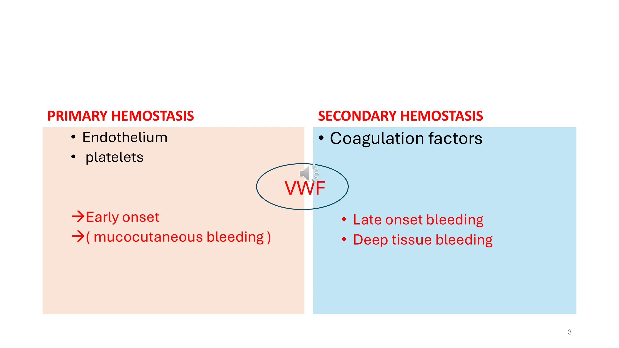
vWF (von Willebrand Factor) bridges primary and secondary hemostasis.
PRIMARY HEMOSTASIS
- Endothelium
- platelets
- → Early onset
- → ( mucocutaneous bleeding )
SECONDARY HEMOSTASIS
- Coagulation factors
- Late onset bleeding
- Deep tissue bleeding
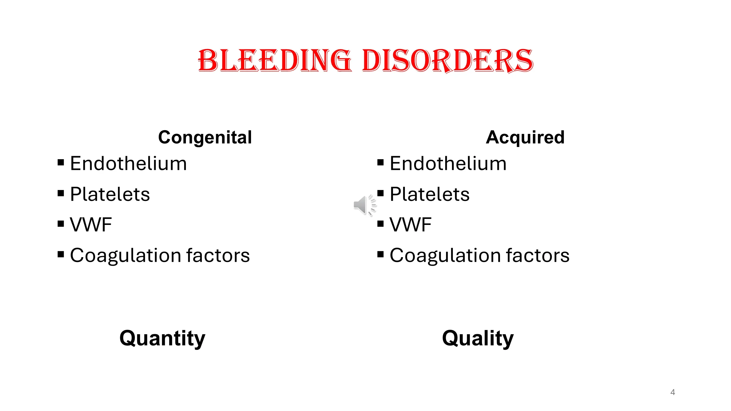
BLEEDING DISORDERS
Congenital
- Endothelium
- Platelets
- VWF
- Coagulation factors
Quantity
Acquired
- Endothelium
- Platelets
- VWF
- Coagulation factors
Quality
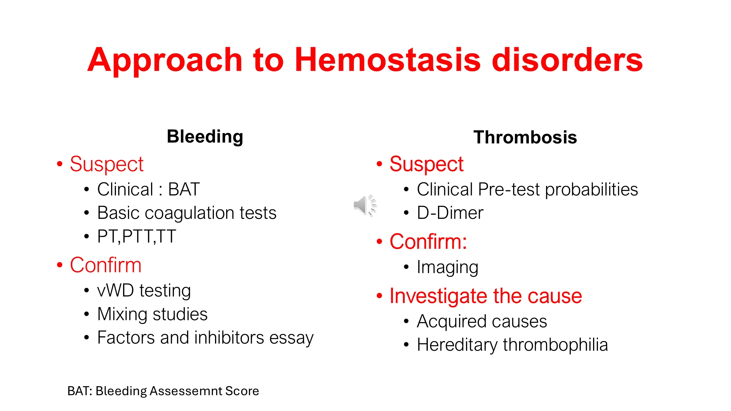
Approach to Hemostasis disorders
Bleeding
- Suspect
- Clinical : BAT
- Basic coagulation tests
- PT,PTT,TT
- Confirm
- vWD testing
- Mixing studies
- Factors and inhibitors essay
Thrombosis
- Suspect
- Clinical Pre-test probabilities
- D-Dimer
- Confirm:
- Imaging
- Investigate the cause
- Acquired causes
- Hereditary thrombophilia
BAT: Bleeding Assessemnt Score
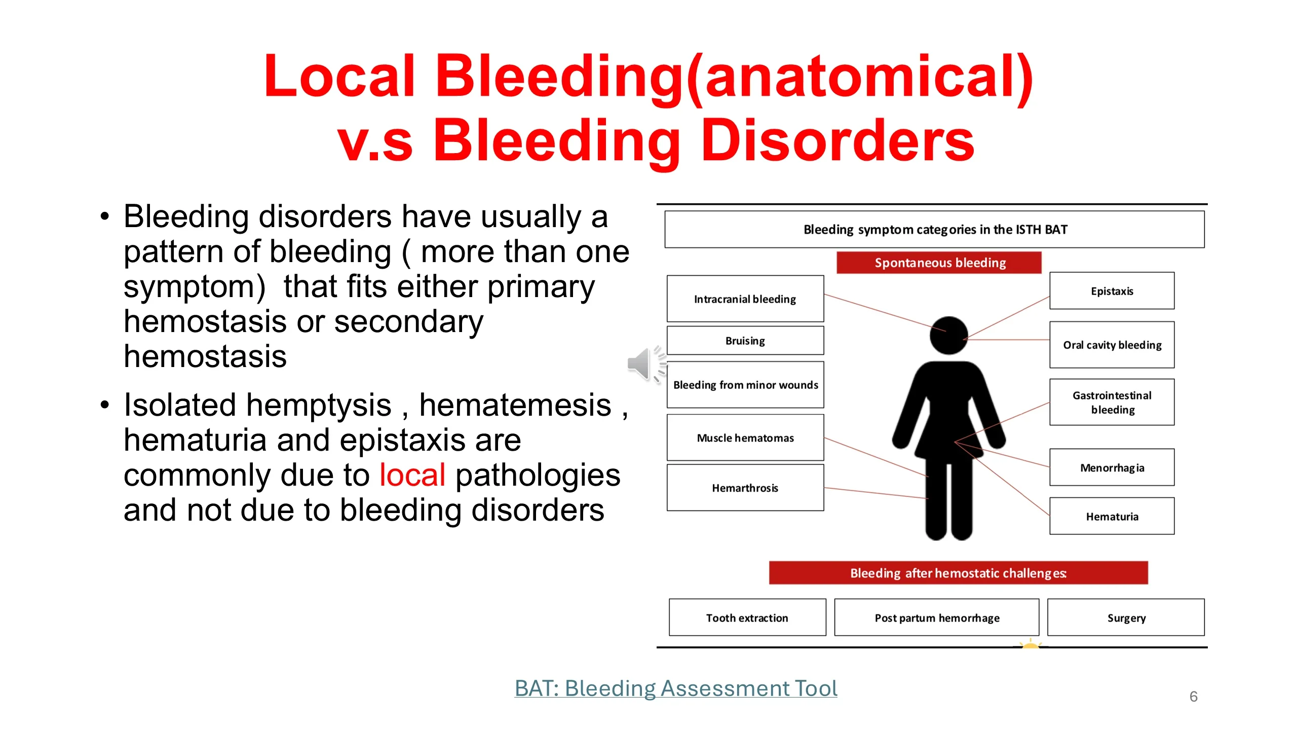
Local Bleeding (anatomical) v.s Bleeding Disorders
- Bleeding disorders have usually a pattern of bleeding ( more than one symptom) that fits either primary hemostasis or secondary hemostasis
- Isolated hemptysis , hematemesis , hematuria and epistaxis are commonly due to local pathologies and not due to bleeding disorders
Diagram – Bleeding symptom categories in the ISTH BAT:
- Spontaneous bleeding: Intracranial bleeding; Bruising; Bleeding from minor wounds; Muscle hematomas; Hemarthrosis; Epistaxis; Oral cavity bleeding; Gastrointestinal bleeding; Menorrhagia; Hematuria
- Bleeding after hemostatic challenges: Tooth extraction; Post partum hemorrhage; Surgery
BAT: Bleeding Assessment Tool
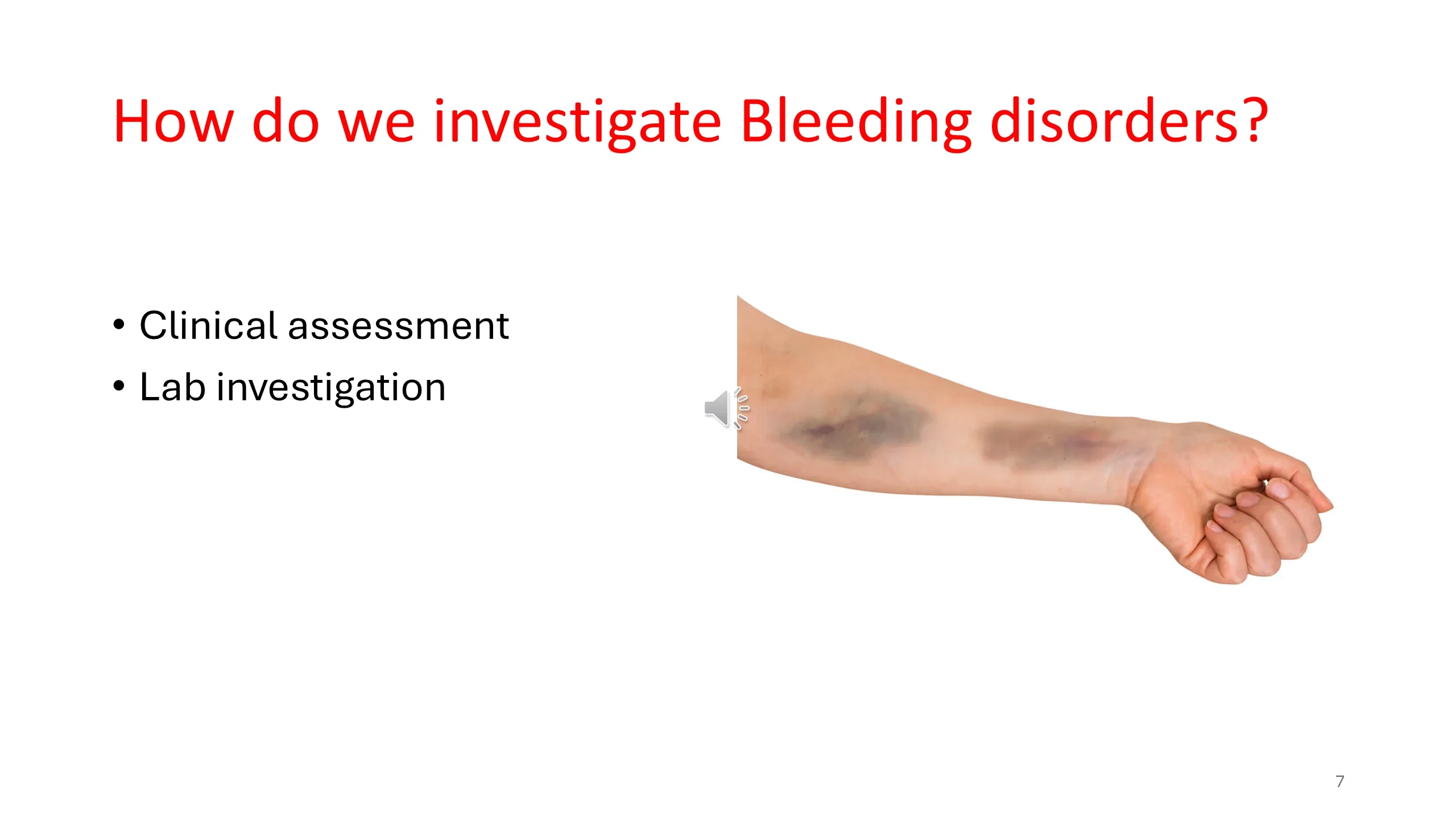
How do we investigate Bleeding disorders?
- Clinical assessment
- Lab investigation
Bleeding : history taking
- HPI and associated symptoms
- Details of the presenting symptom?
- Type of bleeding :
- Muco-cutaneous bleeding (bruising, epistaxis, ICH, GI or GU bleeding,)?
- Deep tissue bleeding : Hemarthrosis, muscle hematomas?
- Other bleeding symptoms , present or past
Suggestive of bleeding disorders
- Bleeding after birth.
- Bleeding after circumcision.
- Large spontaneous bruises, easy gum bleeding, menorrhagia with no gynecological reasons.
- Bleeding after surgery ( dental procedure, appendectomy)
- Family history of bleeding disorders: hemophilia, VWD, hereditary platelets disorders?
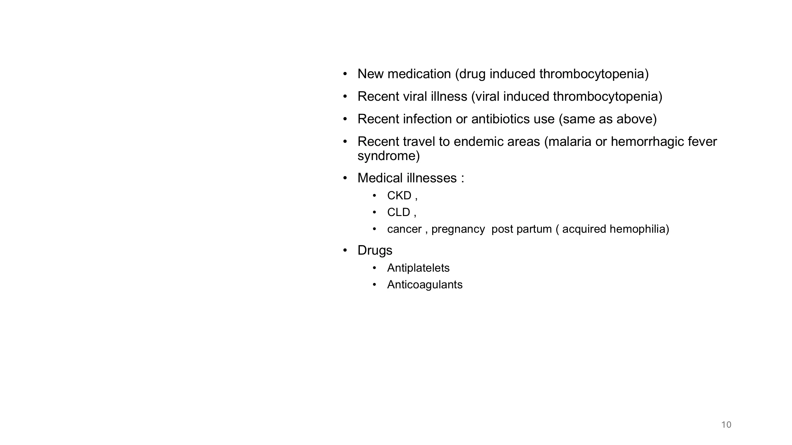
- New medication (drug induced thrombocytopenia)
- Recent viral illness (viral induced thrombocytopenia)
- Recent infection or antibiotics use (same as above)
- Recent travel to endemic areas (malaria or hemorrhagic fever syndrome)
- Medical illnesses :
- CKD ,
- CLD ,
- cancer , pregnancy post partum ( acquired hemophilia)
- Drugs
- Antiplatelets
- Anticoagulants
- Hematology system review
- Does the patient have fatigue, other anemia symptoms or fever ?
- Acquired symptoms of anemia and neutropenia if present along with petechia and mucocutaneus bleeds should alarm you to the presence of pancytopenia
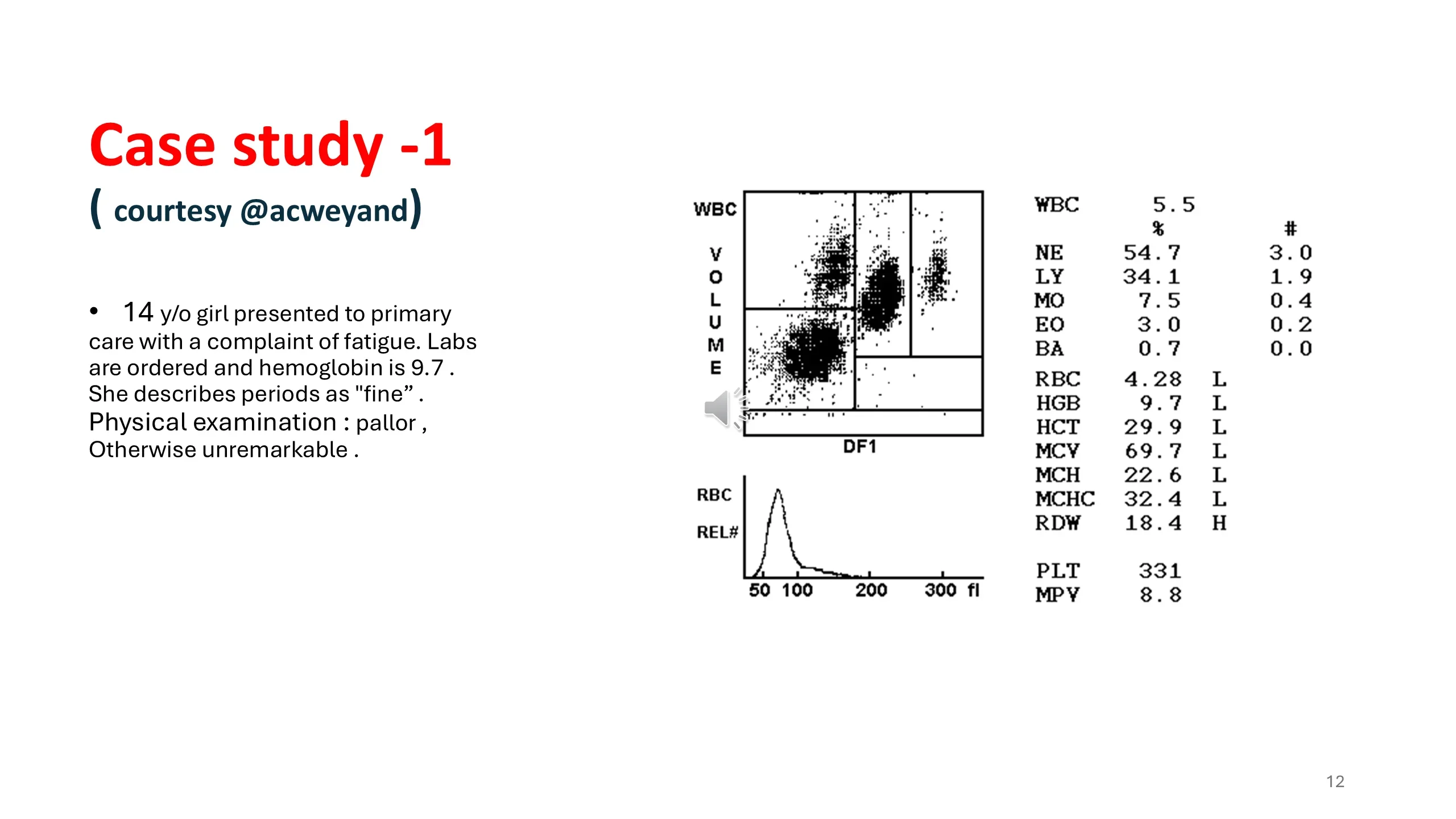
Case study -1 ( courtesy @acweyand)
- 14 y/o girl presented to primary care with a complaint of fatigue. Labs are ordered and hemoglobin is 9.7 . She describes periods as "fine" . Physical examination : pallor , Otherwise unremarkable .
CBC:
| Parameter | % | # | Flag |
|---|---|---|---|
| WBC | 5.5 | ||
| NE | 54.7 | 3.0 | |
| LY | 34.1 | 1.9 | |
| MO | 7.5 | 0.4 | |
| EO | 3.0 | 0.2 | |
| BA | 0.7 | 0.0 | |
| RBC | 4.28 | L | |
| HGB | 9.7 | L | |
| HCT | 29.9 | L | |
| MCV | 69.7 | L | |
| MCH | 22.6 | L | |
| MCHC | 32.4 | L | |
| RDW | 18.4 | H | |
| PLT | 331 | ||
| MPV | 8.8 |
Scattergram axes labeled WBC VOLUME versus DF1; RBC REL# histogram with x-axis 50, 100, 200, 300 fl.
- You decide to delve a little deeper and ask some details. Turns out her "fine" periods last 3-4 weeks!! and she requires pad or tampon changes between every class.
- *key point* many adolescent girls. /women DO NOT KNOW what a normal period should be like.
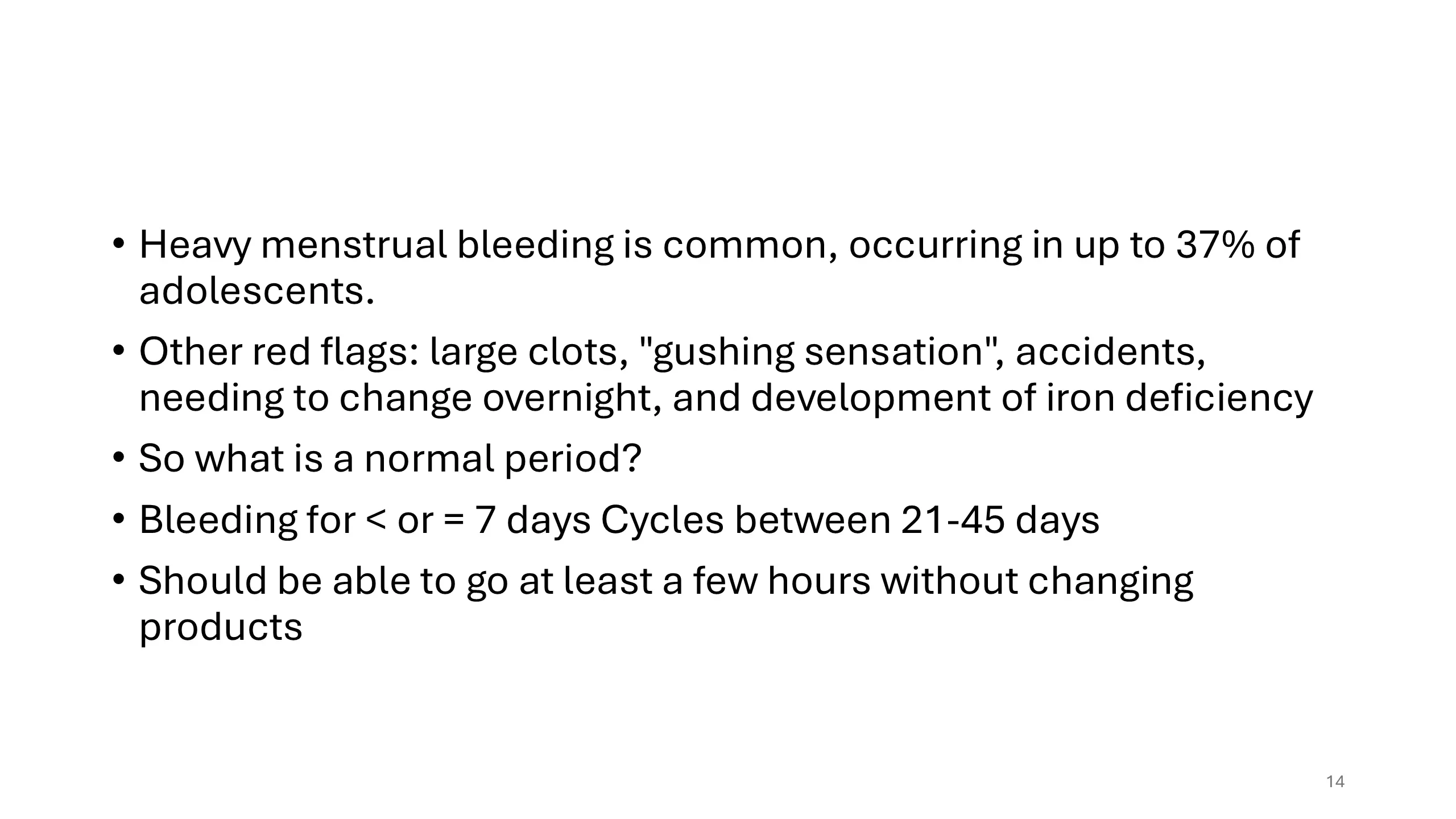
- Heavy menstrual bleeding is common, occurring in up to 37% of adolescents.
- Other red flags: large clots, "gushing sensation", accidents, needing to change overnight, and development of iron deficiency
- So what is a normal period?
- Bleeding for < or = 7 days Cycles between 21-45 days
- Should be able to go at least a few hours without changing products
Heavy menstrual bleeding
- The differential diagnosis for HMB is broad and includes
- Anovulatory cycles (by far the most common),
- coagulopathies (such as VWD)
- STIs, iatrogenic causes,
- Pregnancy.
- Structural causes (very rare in adolescents).
- So what next?
- History should focus on bleeding (epistaxis, oral bleeding, abnormal bruising, surgeries, etc.), medical hx, and family hx.
- BAT
- You decided this patient has significant bleeding history and extra testing is needed.
- How do we investigate bleeding disorders?
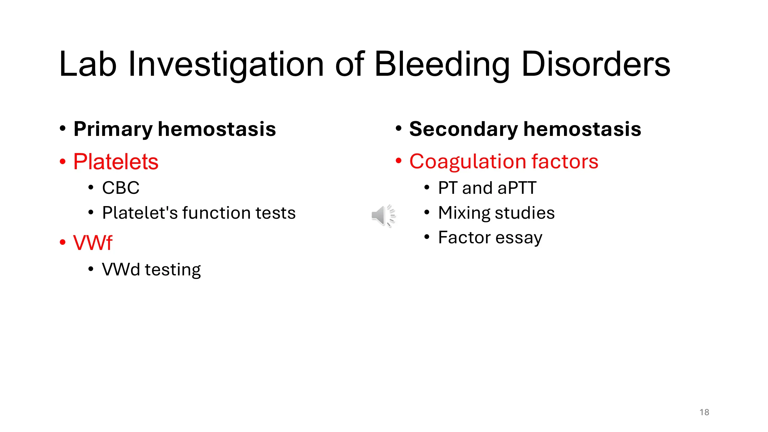
Primary hemostasis
- Platelets
- CBC
- Platelet's function tests
- VWf
- VWd testing
Secondary hemostasis
- Coagulation factors
- PT and aPTT
- Mixing studies
- Factor essay
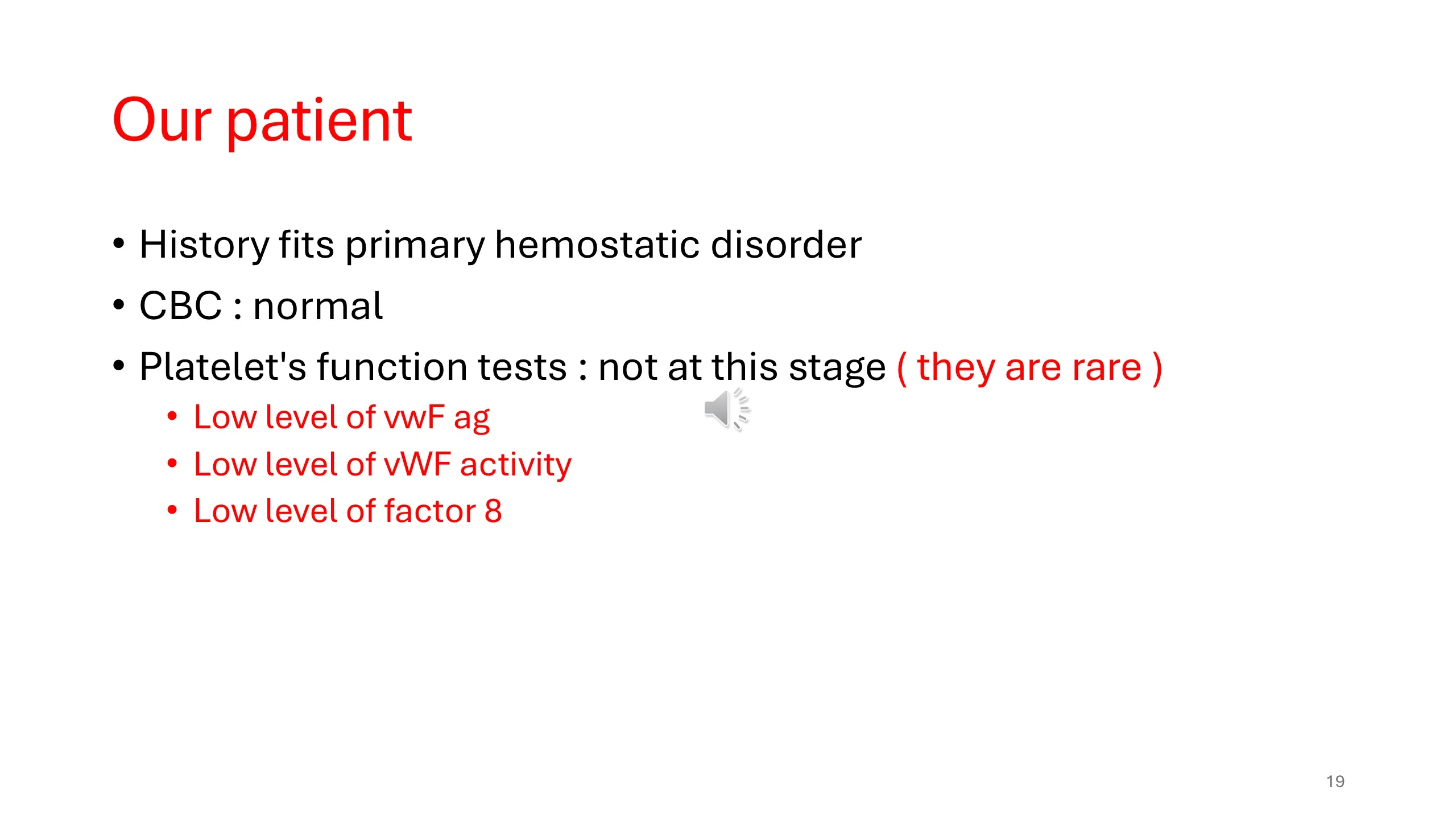
- History fits primary hemostatic disorder
- CBC : normal
- Platelet's function tests : not at this stage ( they are rare )
- Low level of vwF ag
- Low level of vWF activity
- Low level of factor 8
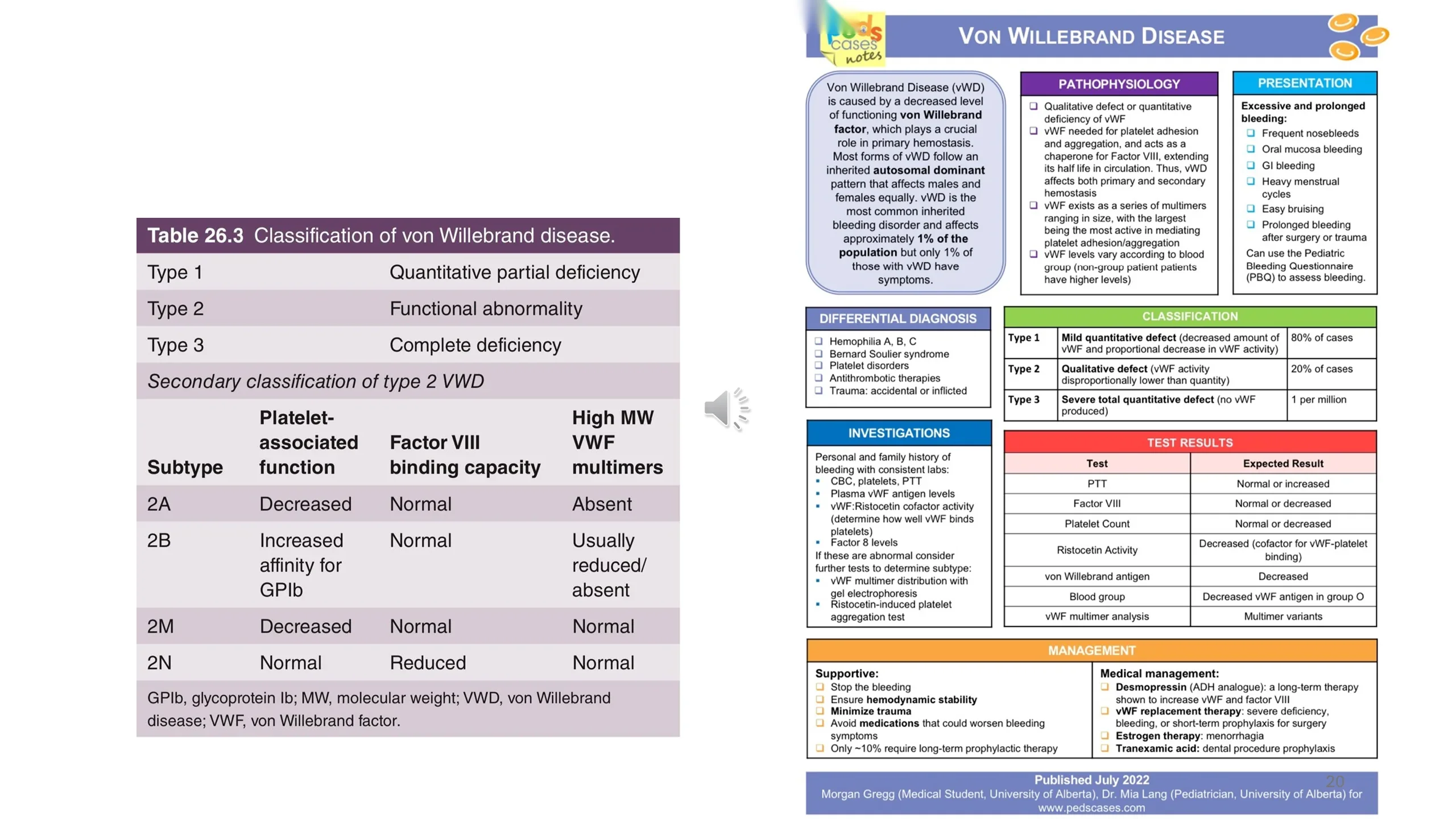
Table 26.3 Classification of von Willebrand disease
| Type | Description |
|---|---|
| Type 1 | Quantitative partial deficiency |
| Type 2 | Functional abnormality |
| Type 3 | Complete deficiency |
Secondary classification of type 2 VWD
| Subtype | Platelet-associated function | Factor VIII binding capacity | High MW VWF multimers |
|---|---|---|---|
| 2A | Decreased | Normal | Absent |
| 2B | Increased affinity for GPIb | Normal | Usually reduced/absent |
| 2M | Decreased | Normal | Normal |
| 2N | Normal | Reduced | Normal |
GPIb, glycoprotein Ib; MW, molecular weight; VWD, von Willebrand disease; VWF, von Willebrand factor.
Von Willebrand Disease (PedsCases Notes summary)
Von Willebrand Disease (vWD) is caused by a decreased level of functioning von Willebrand factor, which plays a crucial role in primary hemostasis. Most forms of vWD follow an inherited autosomal dominant pattern that affects males and females equally. vWD is the most common inherited bleeding disorder and affects approximately 1% of the population but only 1% of those with vWD have symptoms.
Pathophysiology
- Qualitative defect or quantitative deficiency of vWF
- vWF needed for platelet adhesion and aggregation, and acts as a chaperone for Factor VIII, extending its half life in circulation. Thus, vWD affects both primary and secondary hemostasis
- vWF exists as a series of multimers ranging in size, with the largest being the most active in mediating platelet adhesion/aggregation
- vWF levels vary according to blood group (non-group patient patients have higher levels)
Presentation
Excessive and prolonged bleeding:
- Frequent nosebleeds
- Oral mucosa bleeding
- GI bleeding
- Heavy menstrual cycles
- Easy bruising
- Prolonged bleeding after surgery or trauma
Can use the Pediatric Bleeding Questionnaire (PBQ) to assess bleeding.
Differential Diagnosis
- Hemophilia A, B, C
- Bernard Soulier syndrome
- Platelet disorders
- Antithrombotic therapies
- Trauma: accidental or inflicted
Classification
| Type | Description | Frequency |
|---|---|---|
| Type 1 | Mild quantitative defect (decreased amount of vWF and proportional decrease in vWF activity) | 80% of cases |
| Type 2 | Qualitative defect (vWF activity disproportionally lower than quantity) | 20% of cases |
| Type 3 | Severe total quantitative defect (no vWF produced) | 1 per million |
Investigations
Personal and family history of bleeding with consistent labs:
- CBC, platelets, PTT
- Plasma vWF antigen levels
- vWF:Ristocetin cofactor activity (determine how well vWF binds platelets)
- Factor 8 levels
If these are abnormal consider further tests to determine subtype:
- vWF multimer distribution with gel electrophoresis
- Ristocetin-induced platelet aggregation test
Test Results
| Test | Expected Result |
|---|---|
| PTT | Normal or increased |
| Factor VIII | Normal or decreased |
| Platelet Count | Normal or decreased |
| Ristocetin Activity | Decreased (cofactor for vWF-platelet binding) |
| von Willebrand antigen | Decreased |
| Blood group | Decreased vWF antigen in group O |
| vWF multimer analysis | Multimer variants |
Management
Supportive:
- Stop the bleeding
- Ensure hemodynamic stability
- Minimize trauma
- Avoid medications that could worsen bleeding symptoms
- Only ~10% require long-term prophylactic therapy
Medical management:
- Desmopressin (ADH analogue): a long-term therapy shown to increase vWF and factor VIII
- vWF replacement therapy: severe deficiency, bleeding, or short-term prophylaxis for surgery
- Estrogen therapy: menorrhagia
- Tranexamic acid: dental procedure prophylaxis
Published July 2022. Morgan Gregg (Medical Student, University of Alberta), Dr. Mia Lang (Pediatrician, University of Alberta) for www.pedscases.com
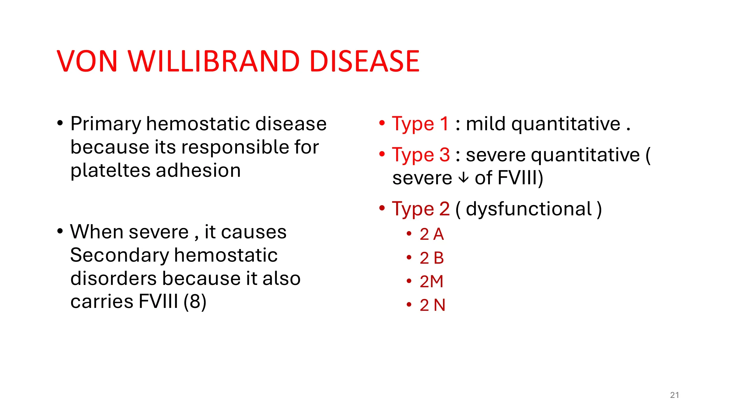
- Primary hemostatic disease because its responsible for plateltes adhesion
- When severe , it causes Secondary hemostatic disorders because it also carries FVIII (8)
- Type 1 : mild quantitative .
- Type 3 : severe quantitative ( severe ↓ of FVIII)
- Type 2 ( dysfunctional )
- 2 A
- 2 B
- 2M
- 2 N
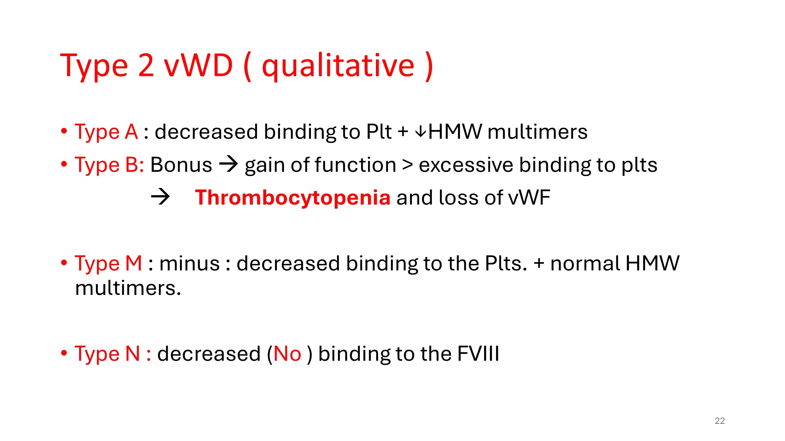
- Type A : decreased binding to Plt + ↓HMW multimers
- Type B: Bonus → gain of function > excessive binding to plts → Thrombocytopenia and loss of vWF
- Type M : minus : decreased binding to the Plts. + normal HMW multimers.
- Type N : decreased (No ) binding to the FVIII
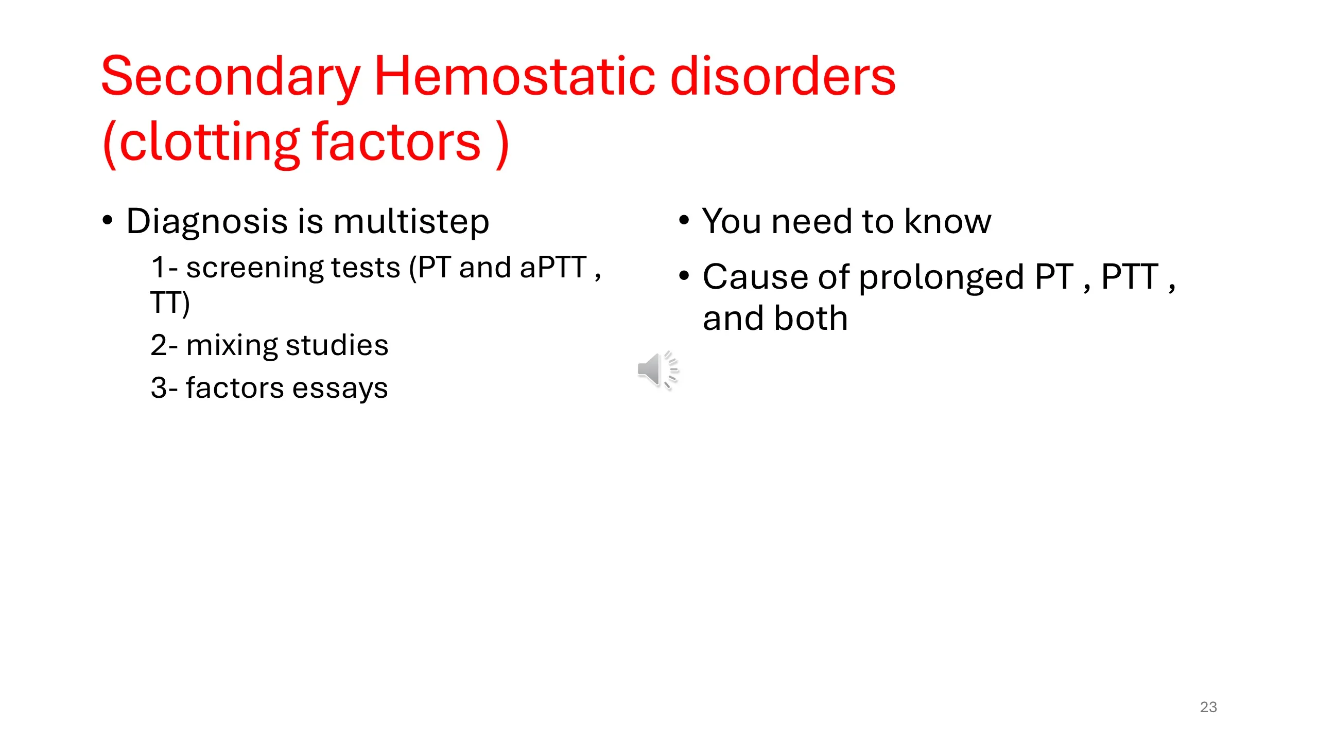
- Diagnosis is multistep
- 1- screening tests (PT and aPTT , TT)
- 2- mixing studies
- 3- factors essays
- You need to know
- Cause of prolonged PT , PTT , and both
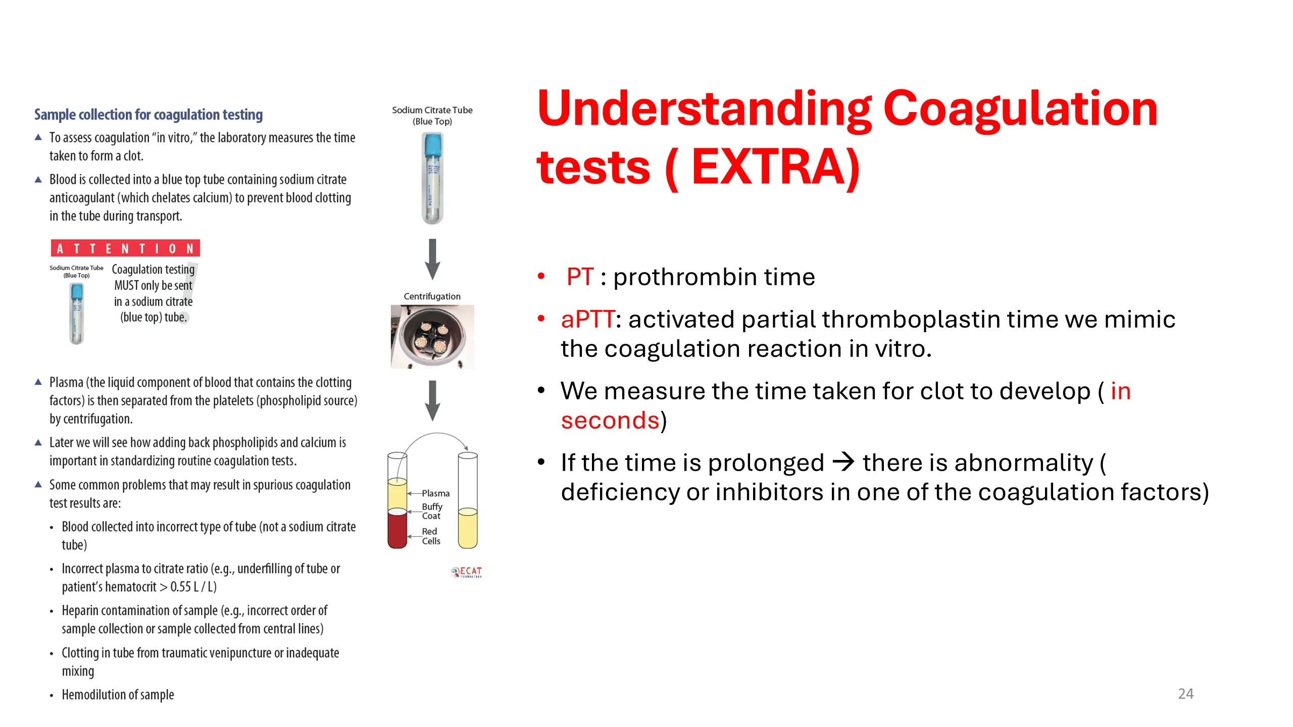
Sample collection for coagulation testing
- To assess coagulation "in vitro," the laboratory measures the time taken to form a clot.
- Blood is collected into a blue top tube containing sodium citrate anticoagulant (which chelates calcium) to prevent blood clotting in the tube during transport.
ATTENTION: Coagulation testing MUST only be sent in a sodium citrate (blue top) tube.
- Plasma (the liquid component of blood that contains the clotting factors) is then separated from the platelets (phospholipid source) by centrifugation.
- Later we will see how adding back phospholipids and calcium is important in standardizing routine coagulation tests.
- Some common problems that may result in spurious coagulation test results are:
- Blood collected into incorrect type of tube (not a sodium citrate tube)
- Incorrect plasma to citrate ratio (e.g., underfilling of tube or patient's hematocrit > 0.55 L / L)
- Heparin contamination of sample (e.g., incorrect order of sample collection or sample collected from central lines)
- Clotting in tube from traumatic venipuncture or inadequate mixing
- Hemodilution of sample
Understanding Coagulation tests ( EXTRA)
- PT : prothrombin time
- aPTT: activated partial thromboplastin time we mimic the coagulation reaction in vitro.
- We measure the time taken for clot to develop ( in seconds)
- If the time is prolonged → there is abnormality ( deficiency or inhibitors in one of the coagulation factors)
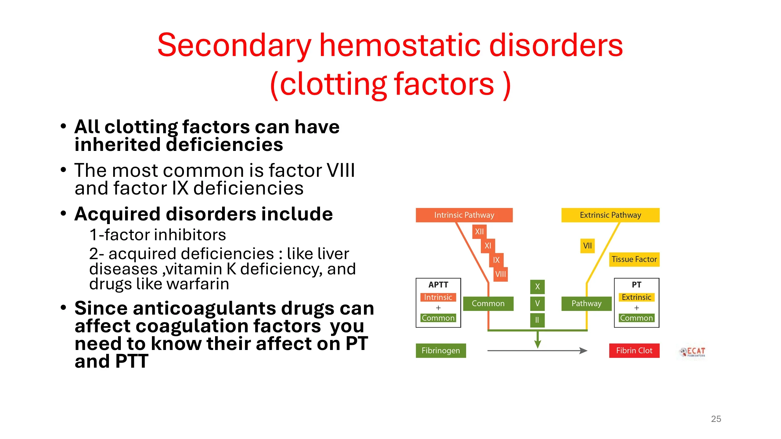
- All clotting factors can have inherited deficiencies
- The most common is factor VIII and factor IX deficiencies
- Acquired disorders include
- 1-factor inhibitors
- 2- acquired deficiencies : like liver diseases, vitamin K deficiency, and drugs like warfarin
- Since anticoagulants drugs can affect coagulation factors you need to know their affect on PT and PTT
Cascade diagram: Intrinsic Pathway (Factors XII, XI, IX, VIII) measured by APTT (intrinsic + common); Extrinsic Pathway (Factor VII, Tissue Factor) measured by PT (extrinsic + common); both feed the Common Pathway (Factors X, V, II) leading to Fibrinogen → Fibrin Clot.
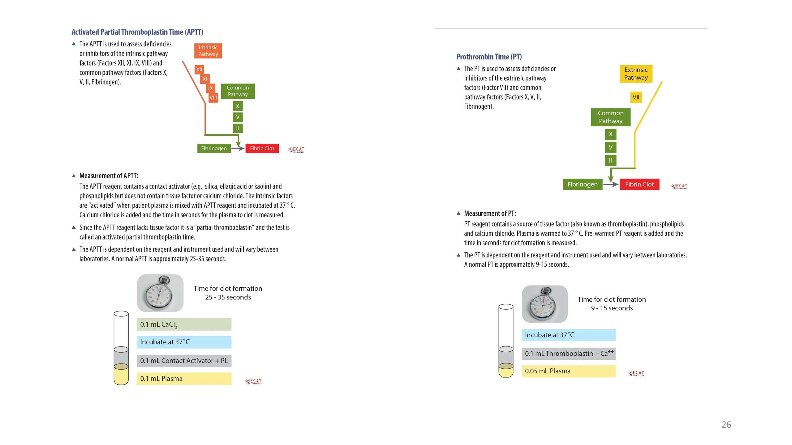
Activated Partial Thromboplastin Time (APTT)
- The APTT is used to assess deficiencies or inhibitors of the intrinsic pathway factors (Factors XII, XI, IX, VIII) and common pathway factors (Factors X, V, II, Fibrinogen).
Cascade: Intrinsic Pathway (XII, XI, IX, VIII) → Common Pathway (X, V, II) → Fibrinogen → Fibrin Clot.
Measurement of APTT:
- The APTT reagent contains a contact activator (e.g., silica, ellagic acid or kaolin) and phospholipids but does not contain tissue factor or calcium chloride. The intrinsic factors are "activated" when patient plasma is mixed with APTT reagent and incubated at 37 °C. Calcium chloride is added and the time in seconds for the plasma to clot is measured.
- Since the APTT reagent lacks tissue factor it is a "partial thromboplastin" and the test is called an activated partial thromboplastin time.
- The APTT is dependent on the reagent and instrument used and will vary between laboratories. A normal APTT is approximately 25-35 seconds.
Reaction tube (bottom to top): 0.1 mL Plasma; 0.1 mL Contact Activator + PL; Incubate at 37°C; 0.1 mL CaCl2. Time for clot formation 25 - 35 seconds.
Prothrombin Time (PT)
- The PT is used to assess deficiencies or inhibitors of the extrinsic pathway factors (Factor VII) and common pathway factors (Factors X, V, II, Fibrinogen).
Cascade: Extrinsic Pathway (VII) → Common Pathway (X, V, II) → Fibrinogen → Fibrin Clot.
Measurement of PT:
- PT reagent contains a source of tissue factor (also known as thromboplastin), phospholipids and calcium chloride. Plasma is warmed to 37°C. Pre-warmed PT reagent is added and the time in seconds for clot formation is measured.
- The PT is dependent on the reagent and instrument used and will vary between laboratories. A normal PT is approximately 9-15 seconds.
Reaction tube (bottom to top): 0.05 mL Plasma; 0.1 mL Thromboplastin + Ca++; Incubate at 37°C. Time for clot formation 9 - 15 seconds.
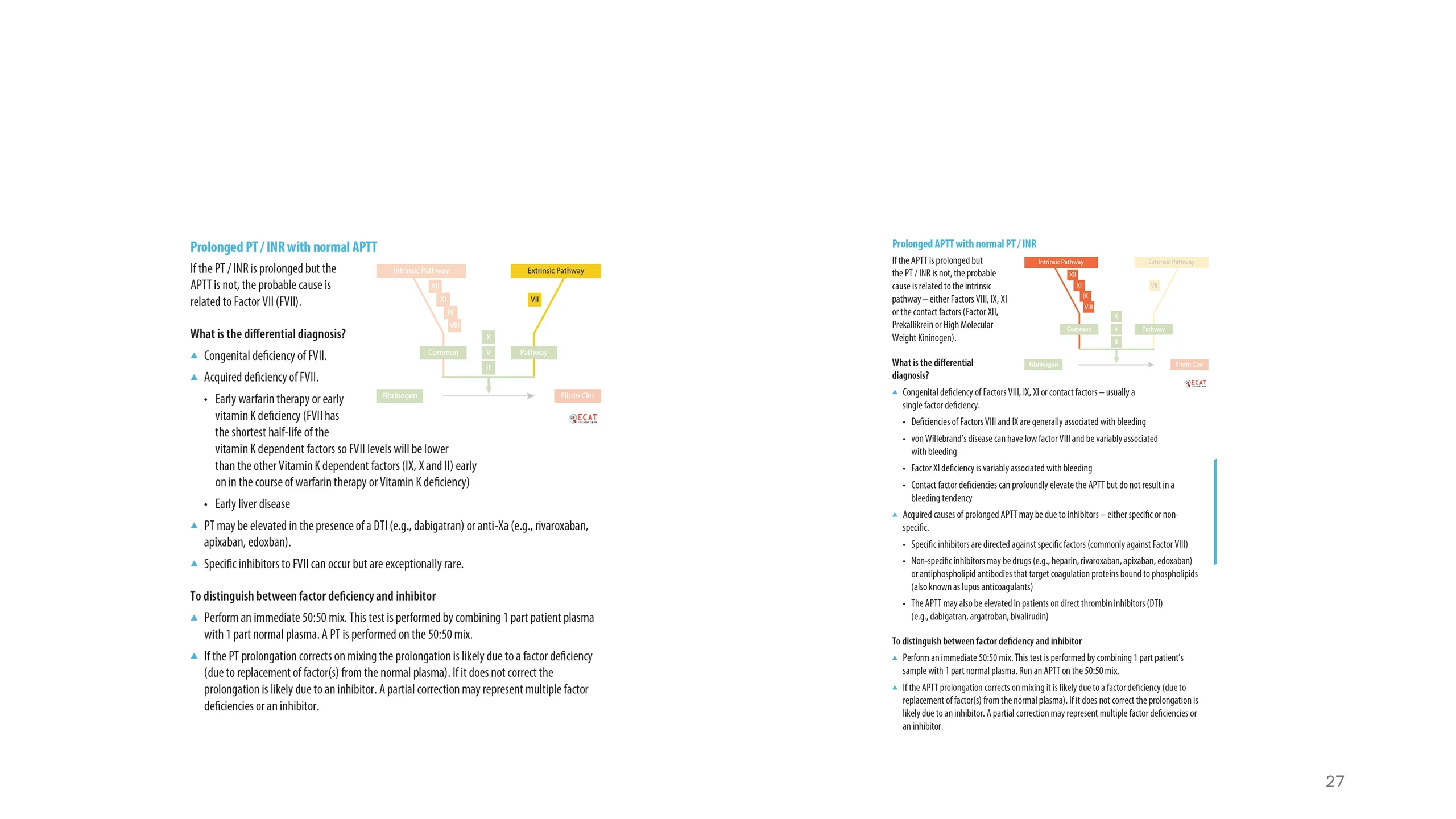
Prolonged PT / INR with normal APTT
If the PT / INR is prolonged but the APTT is not, the probable cause is related to Factor VII (FVII).
What is the differential diagnosis?
- Congenital deficiency of FVII.
- Acquired deficiency of FVII.
- Early warfarin therapy or early vitamin K deficiency (FVII has the shortest half-life of the vitamin K dependent factors so FVII levels will be lower than the other Vitamin K dependent factors (IX, X and II) early on in the course of warfarin therapy or Vitamin K deficiency)
- Early liver disease
- PT may be elevated in the presence of a DTI (e.g., dabigatran) or anti-Xa (e.g., rivaroxaban, apixaban, edoxban).
- Specific inhibitors to FVII can occur but are exceptionally rare.
To distinguish between factor deficiency and inhibitor
- Perform an immediate 50:50 mix. This test is performed by combining 1 part patient plasma with 1 part normal plasma. A PT is performed on the 50:50 mix.
- If the PT prolongation corrects on mixing the prolongation is likely due to a factor deficiency (due to replacement of factor(s) from the normal plasma). If it does not correct the prolongation is likely due to an inhibitor. A partial correction may represent multiple factor deficiencies or an inhibitor.
Prolonged APTT with normal PT / INR
If the APTT is prolonged but the PT / INR is not, the probable cause is related to the intrinsic pathway – either Factors VIII, IX, XI or the contact factors (Factor XII, Prekallikrein or High Molecular Weight Kininogen).
What is the differential diagnosis?
- Congenital deficiency of Factors VIII, IX, XI or contact factors – usually a single factor deficiency.
- Deficiencies of Factors VIII and IX are generally associated with bleeding
- von Willebrand's disease can have low factor VIII and be variably associated with bleeding
- Factor XI deficiency is variably associated with bleeding
- Contact factor deficiencies can profoundly elevate the APTT but do not result in a bleeding tendency
- Acquired causes of prolonged APTT may be due to inhibitors – either specific or non-specific.
- Specific inhibitors are directed against specific factors (commonly against Factor VIII)
- Non-specific inhibitors may be drugs (e.g., heparin, rivaroxaban, apixaban, edoxaban) or antiphospholipid antibodies that target coagulation proteins bound to phospholipids (also known as lupus anticoagulants)
- The APTT may also be elevated in patients on direct thrombin inhibitors (DTI) (e.g., dabigatran, argatroban, bivalirudin)
To distinguish between factor deficiency and inhibitor
- Perform an immediate 50:50 mix. This test is performed by combining 1 part patient's sample with 1 part normal plasma. Run an APTT on the 50:50 mix.
- If the APTT prolongation corrects on mixing it is likely due to a factor deficiency (due to replacement of factor(s) from the normal plasma). If it does not correct the prolongation is likely due to an inhibitor. A partial correction may represent multiple factor deficiencies or an inhibitor.
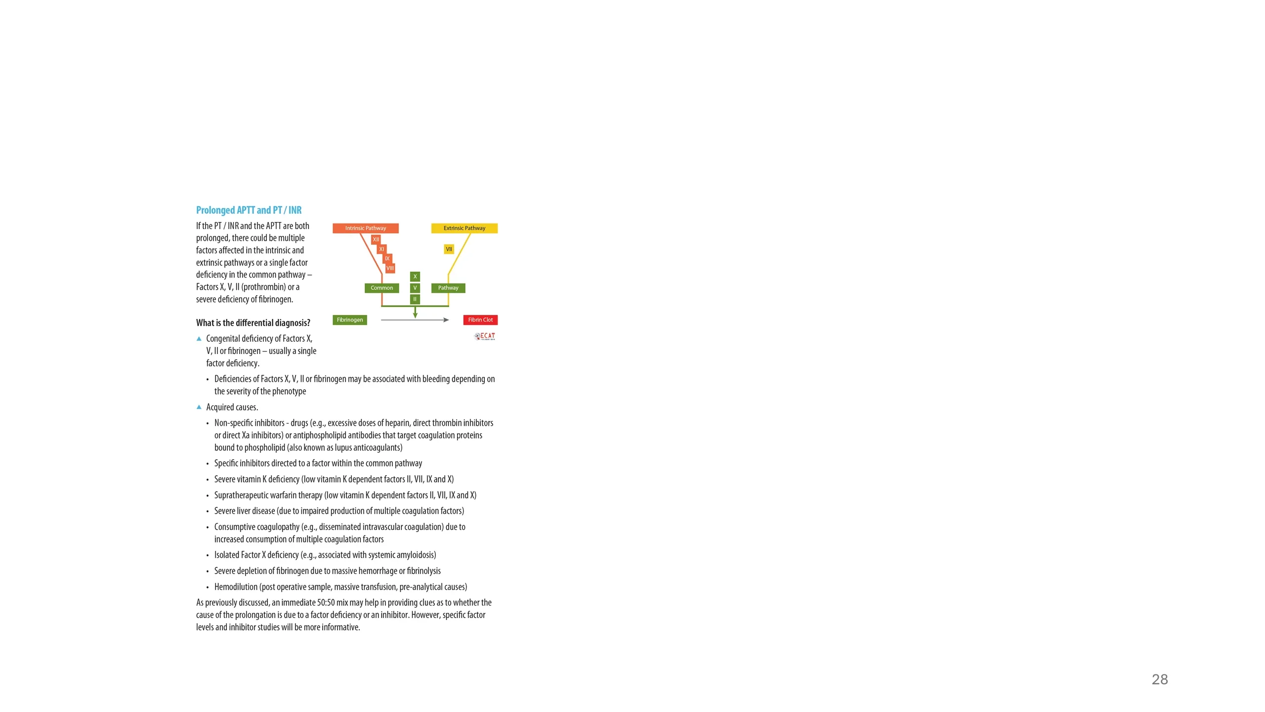
Prolonged APTT and PT / INR
If the PT / INR and the APTT are both prolonged, there could be multiple factors affected in the intrinsic and extrinsic pathways or a single factor deficiency in the common pathway – Factors X, V, II (prothrombin) or a severe deficiency of fibrinogen.
What is the differential diagnosis?
- Congenital deficiency of Factors X, V, II or fibrinogen – usually a single factor deficiency.
- Deficiencies of Factors X, V, II or fibrinogen may be associated with bleeding depending on the severity of the phenotype
- Acquired causes.
- Non-specific inhibitors - drugs (e.g., excessive doses of heparin, direct thrombin inhibitors or direct Xa inhibitors) or antiphospholipid antibodies that target coagulation proteins bound to phospholipid (also known as lupus anticoagulants)
- Specific inhibitors directed to a factor within the common pathway
- Severe vitamin K deficiency (low vitamin K dependent factors II, VII, IX and X)
- Supratherapeutic warfarin therapy (low vitamin K dependent factors II, VII, IX and X)
- Severe liver disease (due to impaired production of multiple coagulation factors)
- Consumptive coagulopathy (e.g., disseminated intravascular coagulation) due to increased consumption of multiple coagulation factors
- Isolated Factor X deficiency (e.g., associated with systemic amyloidosis)
- Severe depletion of fibrinogen due to massive hemorrhage or fibrinolysis
- Hemodilution (post operative sample, massive transfusion, pre-analytical causes)
As previously discussed, an immediate 50:50 mix may help in providing clues as to whether the cause of the prolongation is due to a factor deficiency or an inhibitor. However, specific factor levels and inhibitor studies will be more informative.
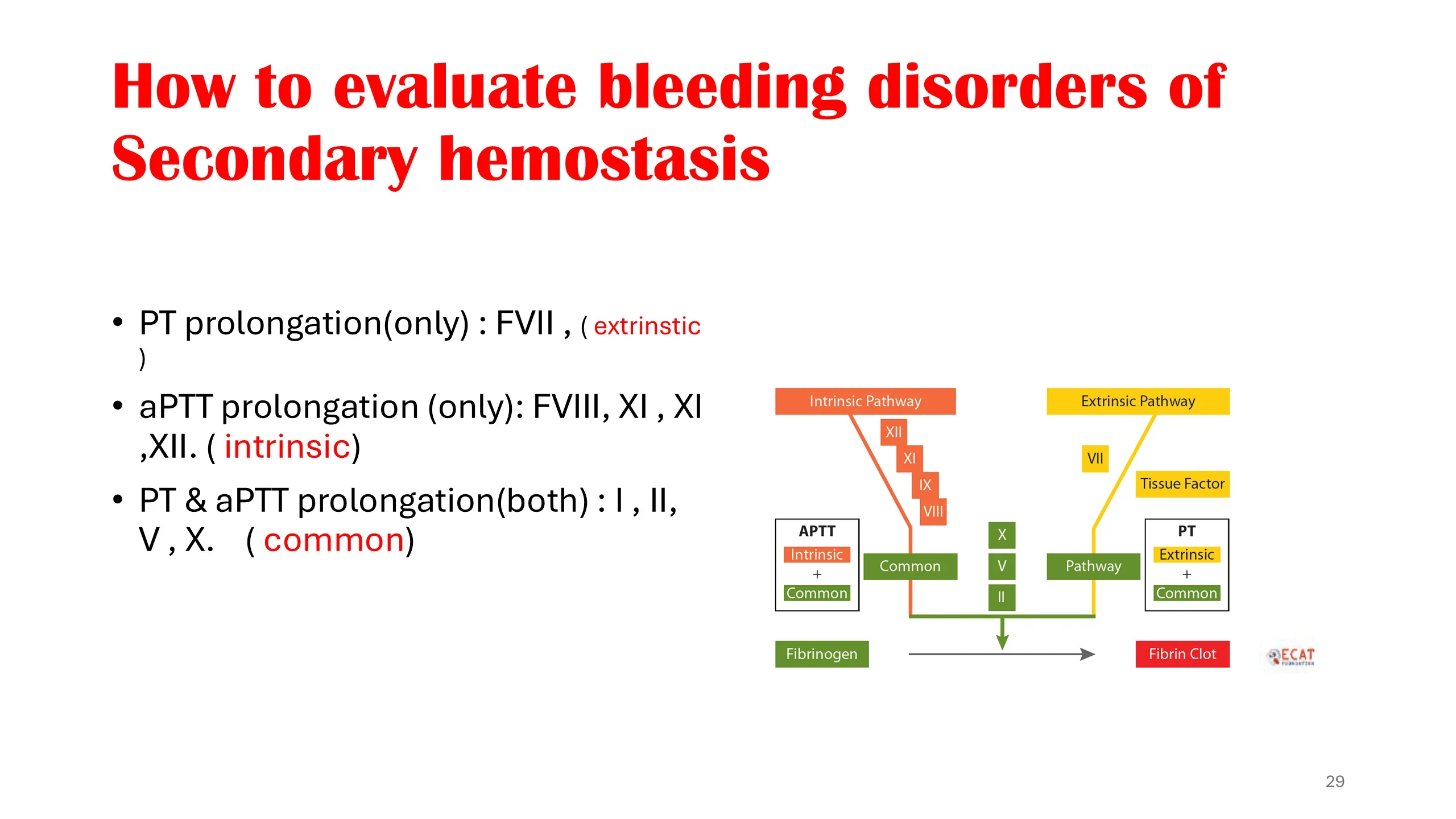
- PT prolongation(only) : FVII , ( extrinstic )
- aPTT prolongation (only): FVIII, XI , XI ,XII. ( intrinsic)
- PT & aPTT prolongation(both) : I , II, V , X. ( common)
Cascade diagram: Intrinsic Pathway (XII, XI, IX, VIII) and Common (measured by APTT: Intrinsic + Common); Extrinsic Pathway (VII, Tissue Factor) measured by PT (Extrinsic + Common); Common Pathway (X, V, II) → Fibrinogen → Fibrin Clot.
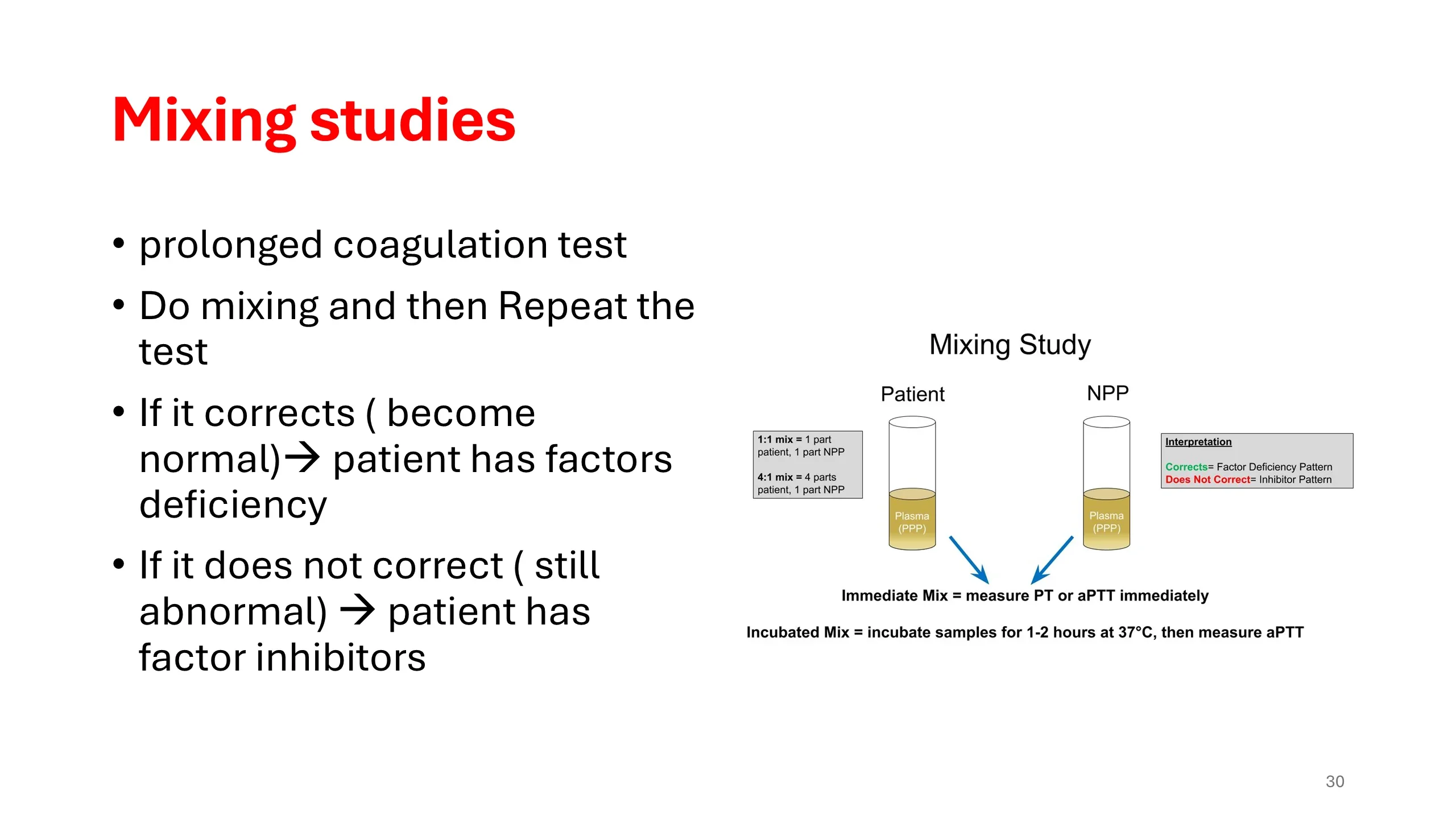
- prolonged coagulation test
- Do mixing and then Repeat the test
- If it corrects ( become normal) → patient has factors deficiency
- If it does not correct ( still abnormal) → patient has factor inhibitors
Mixing Study diagram: mix Patient plasma with NPP. 1:1 mix = 1 part patient, 1 part NPP; 4:1 mix = 4 parts patient, 1 part NPP. Plasma used is PPP. Immediate Mix = measure PT or aPTT immediately. Incubated Mix = incubate samples for 1-2 hours at 37°C, then measure aPTT. Interpretation: Corrects = Factor Deficiency Pattern; Does Not Correct = Inhibitor Pattern.
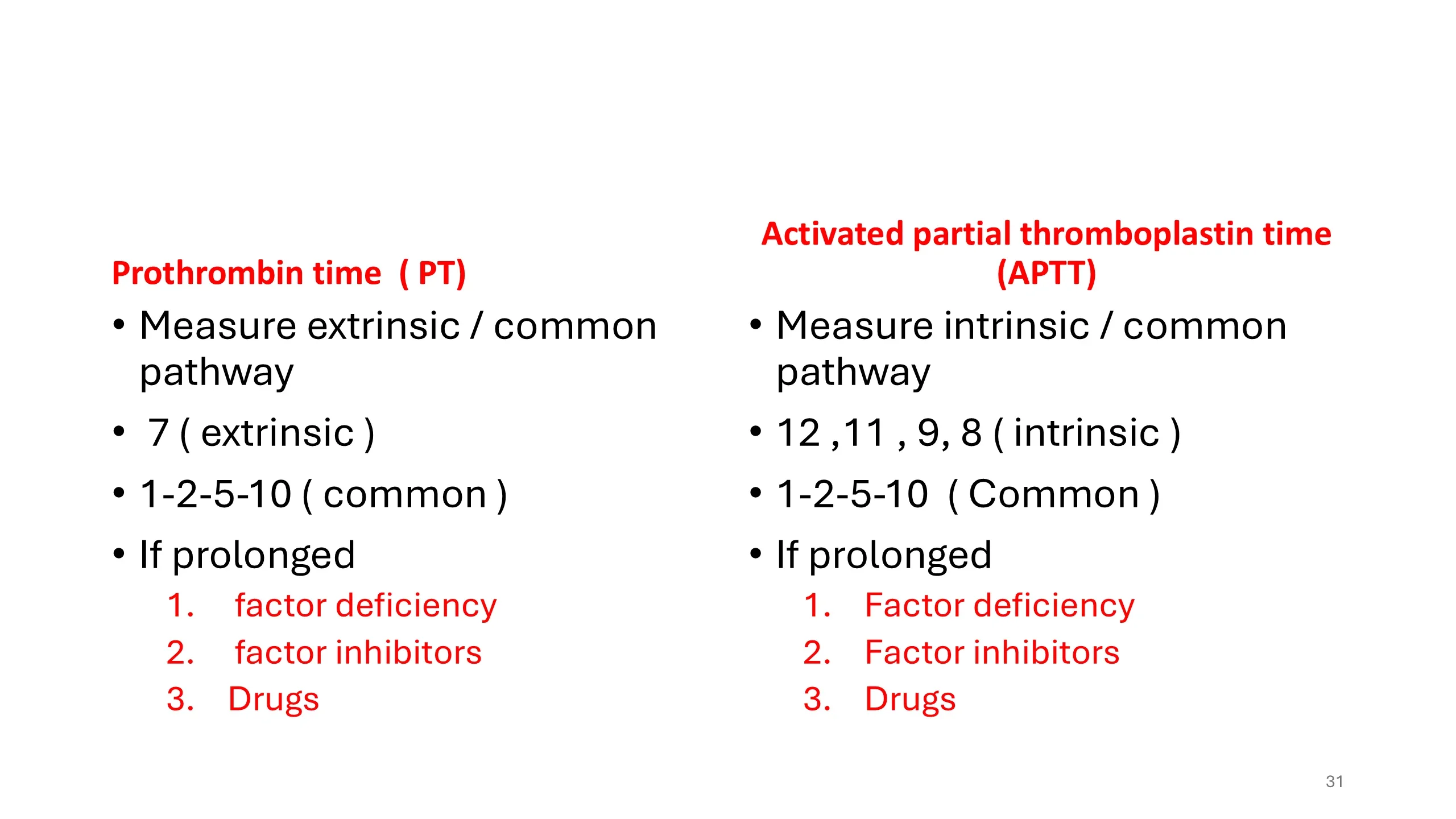
Prothrombin time ( PT)
- Measure extrinsic / common pathway
- 7 ( extrinsic )
- 1-2-5-10 ( common )
- If prolonged
- factor deficiency
- factor inhibitors
- Drugs
Activated partial thromboplastin time (APTT)
- Measure intrinsic / common pathway
- 12 ,11 , 9, 8 ( intrinsic )
- 1-2-5-10 ( Common )
- If prolonged
- Factor deficiency
- Factor inhibitors
- Drugs
Reference
- For the exam
- bleeding lecture
- Thrombosis seminar
- Any book of your choice
- Suggestions will be shared with you .
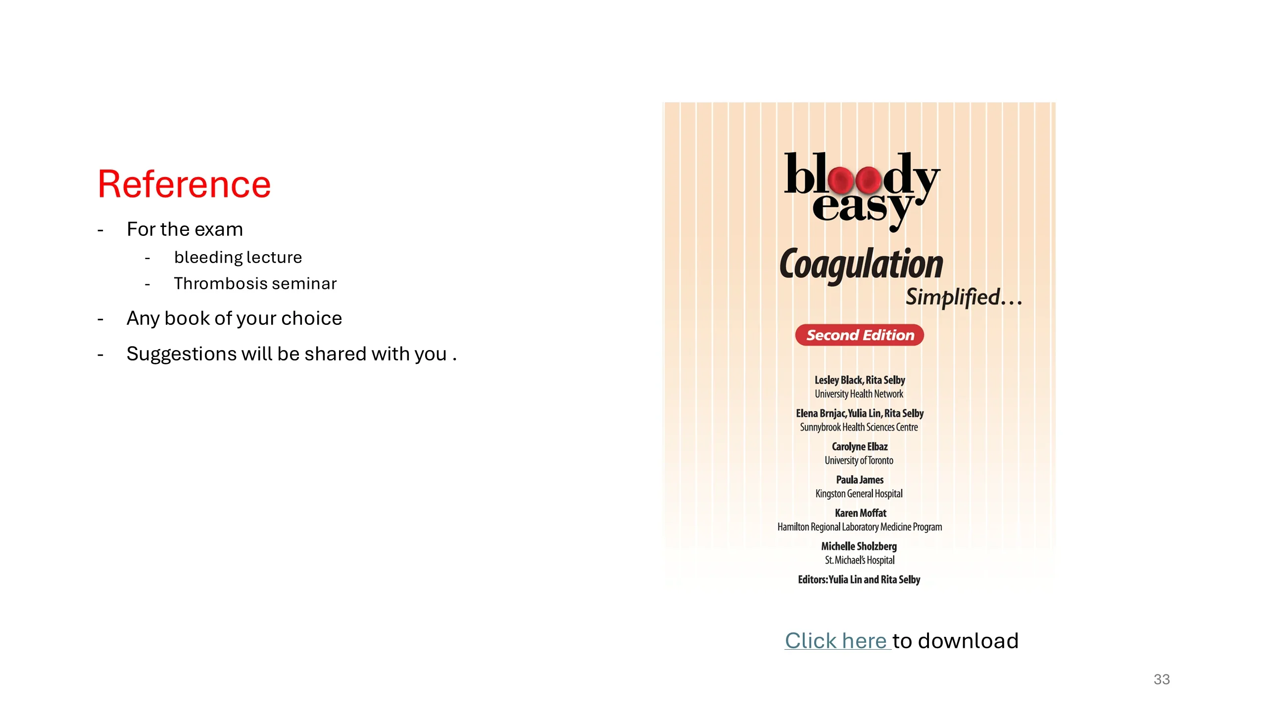
Book cover: Bloody Easy Coagulation Simplified…, Second Edition. Lesley Black, Rita Selby (University Health Network); Elena Brnjac, Yulia Lin, Rita Selby (Sunnybrook Health Sciences Centre); Carolyne Elbaz (University of Toronto); Paula James (Kingston General Hospital); Karen Moffat (Hamilton Regional Laboratory Medicine Program); Michelle Sholzberg (St. Michael's Hospital). Editors: Yulia Lin and Rita Selby.
Click here to download