Acute Leukemia
From origin to emergency — pathogenesis, diagnosis, and the oncologic emergencies.
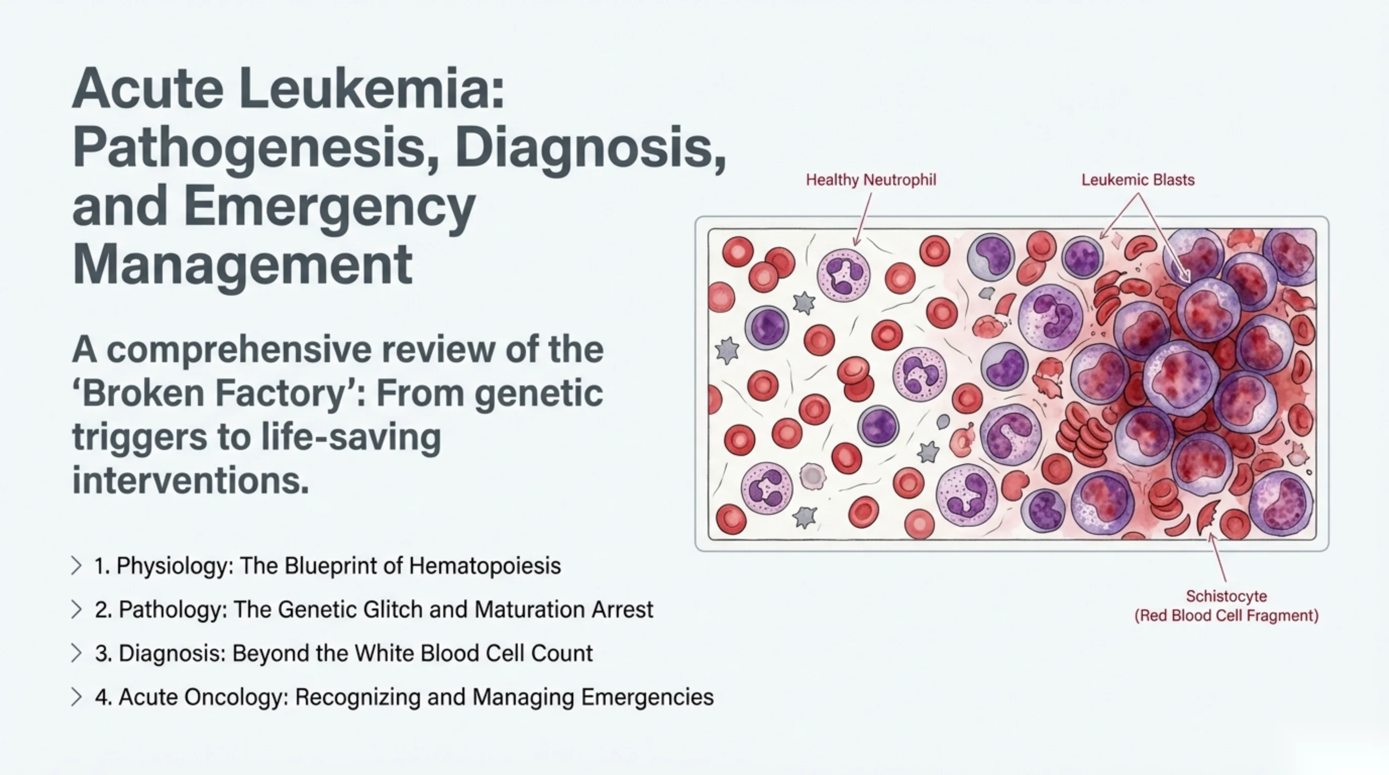
Acute Leukemia: Pathogenesis, Diagnosis, and Emergency Management
A comprehensive review of the ‘Broken Factory’: From genetic triggers to life-saving interventions.
- 1. Physiology: The Blueprint of Hematopoiesis
- 2. Pathology: The Genetic Glitch and Maturation Arrest
- 3. Diagnosis: Beyond the White Blood Cell Count
- 4. Acute Oncology: Recognizing and Managing Emergencies
Illustration labels: Healthy Neutrophil; Leukemic Blasts; Schistocyte (Red Blood Cell Fragment).
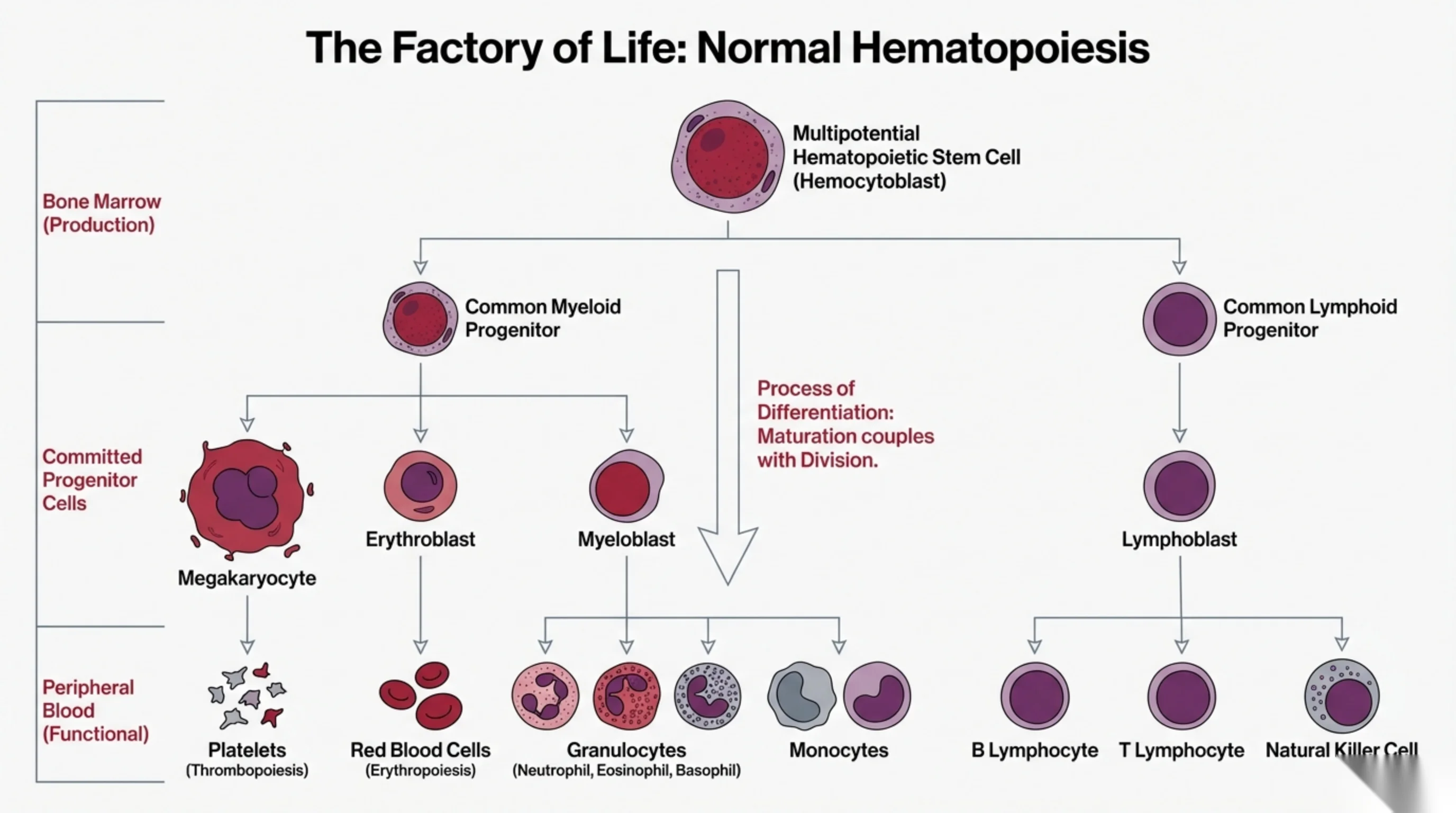
The Factory of Life: Normal Hematopoiesis
The chart is organized into three anatomical/functional tiers on the left: Bone Marrow (Production); Committed Progenitor Cells; and Peripheral Blood (Functional).
Differentiation flow:
- Multipotential Hematopoietic Stem Cell (Hemocytoblast) → gives rise to the Common Myeloid Progenitor and the Common Lymphoid Progenitor.
- Common Myeloid Progenitor → Megakaryocyte, Erythroblast, and Myeloblast.
- Megakaryocyte → Platelets (Thrombopoiesis).
- Erythroblast → Red Blood Cells (Erythropoiesis).
- Myeloblast → Granulocytes (Neutrophil, Eosinophil, Basophil) and Monocytes.
- Common Lymphoid Progenitor → Lymphoblast → B Lymphocyte, T Lymphocyte, and Natural Killer Cell.
Annotation on the central maturation arrow: “Process of Differentiation: Maturation couples with Division.”
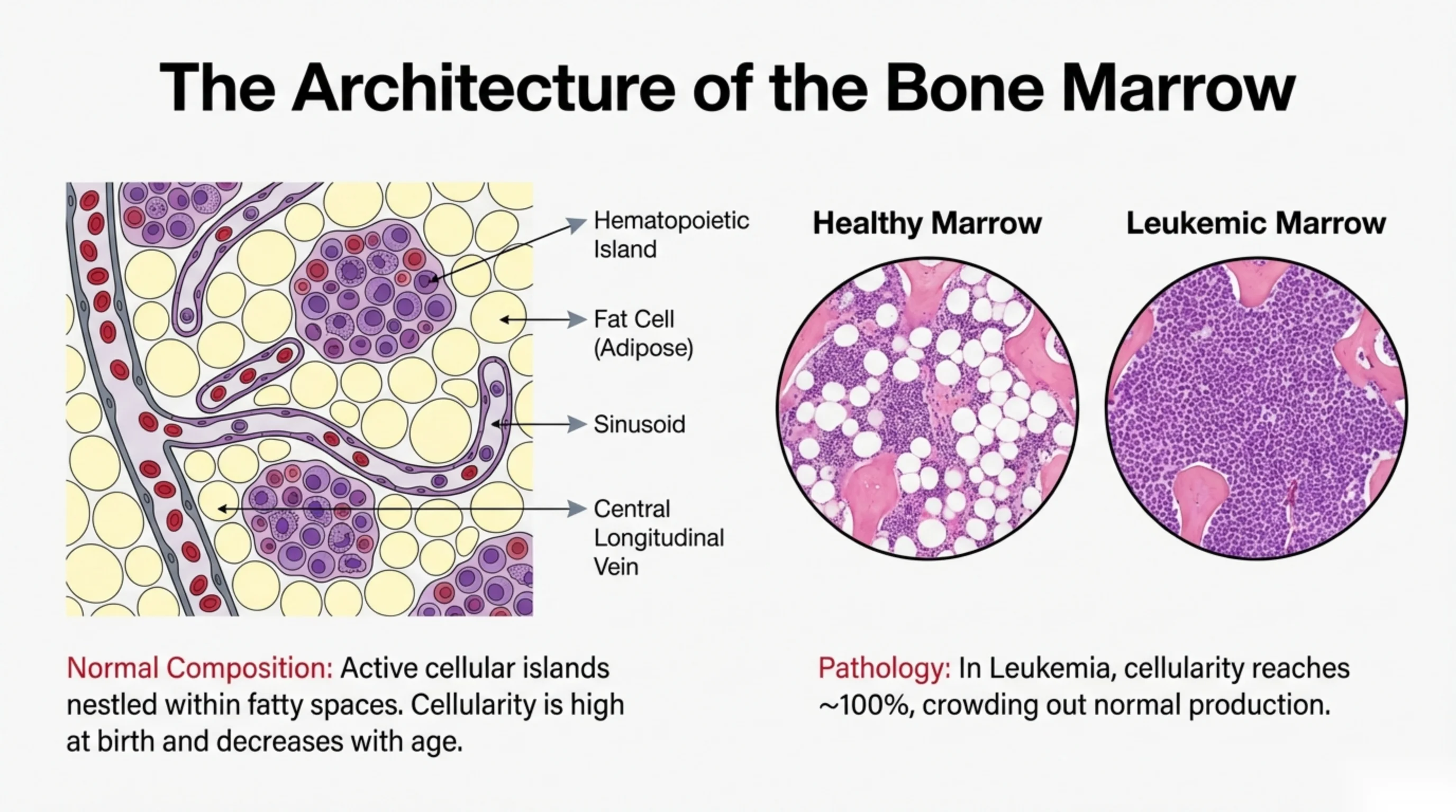
The Architecture of the Bone Marrow
Labeled structures on the marrow illustration: Hematopoietic Island; Fat Cell (Adipose); Sinusoid; Central Longitudinal Vein.
Two comparison micrographs are shown: “Healthy Marrow” and “Leukemic Marrow”.
Normal Composition: Active cellular islands nestled within fatty spaces. Cellularity is high at birth and decreases with age.
Pathology: In Leukemia, cellularity reaches ~100%, crowding out normal production.
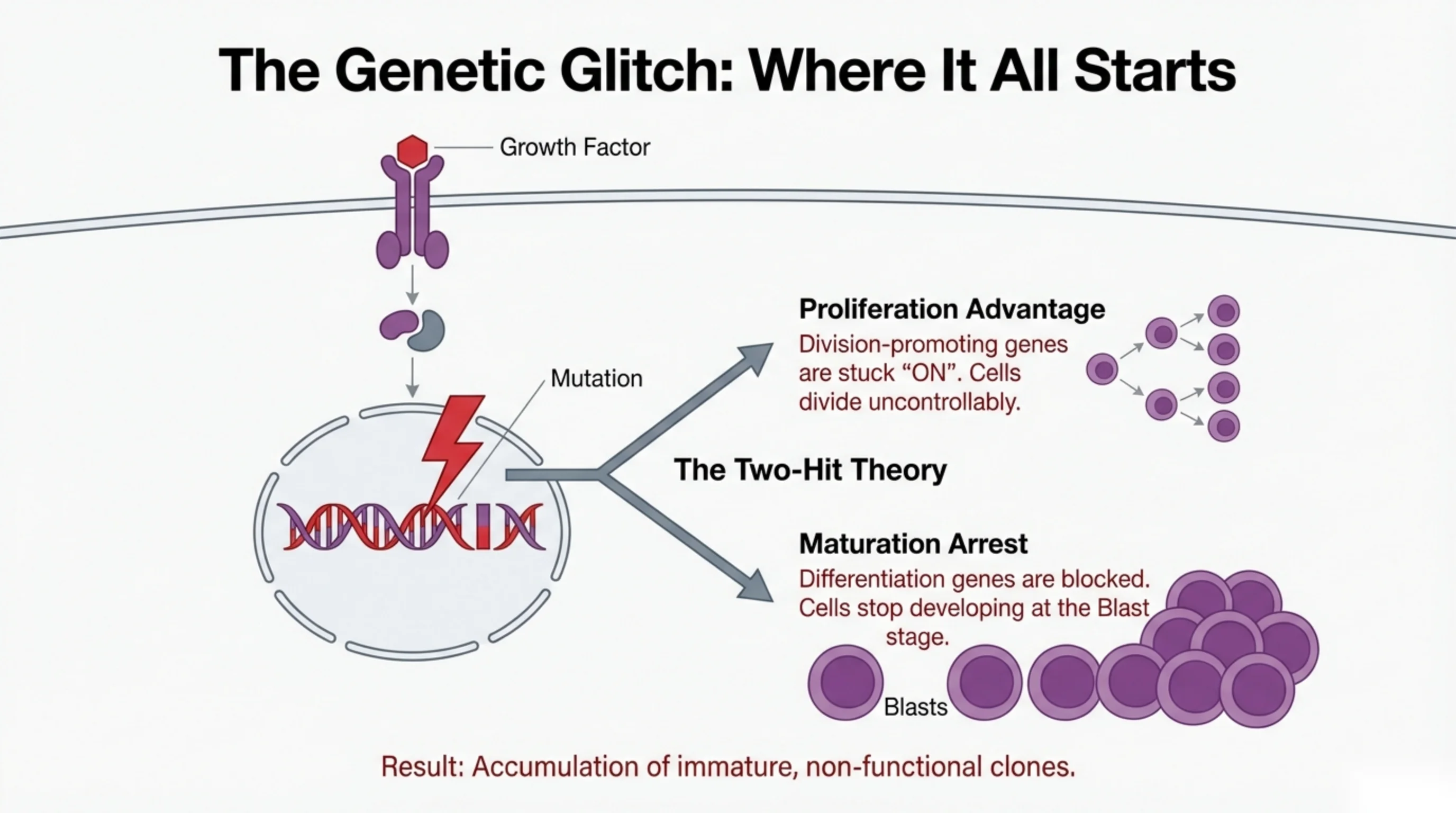
The Genetic Glitch: Where It All Starts
A Growth Factor binds a cell-surface receptor, triggering intracellular signaling to the nucleus, where a Mutation strikes the DNA. This branches into two outcomes (labeled The Two-Hit Theory):
- Proliferation Advantage: Division-promoting genes are stuck “ON”. Cells divide uncontrollably.
- Maturation Arrest: Differentiation genes are blocked. Cells stop developing at the Blast stage. (Illustration labeled “Blasts”.)
Result: Accumulation of immature, non-functional clones.
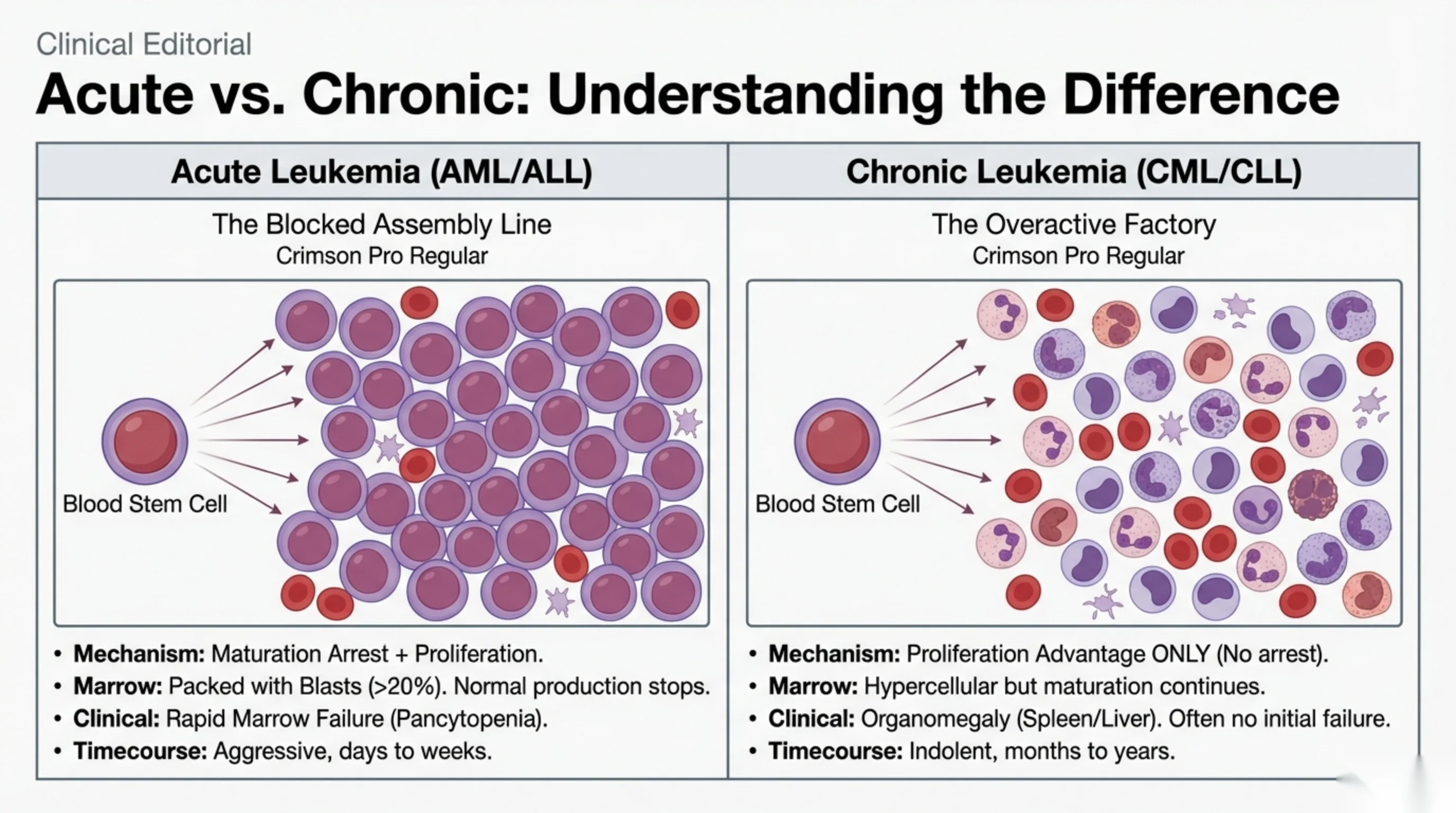
Clinical Editorial
Acute vs. Chronic: Understanding the Difference
| Acute Leukemia (AML/ALL) | Chronic Leukemia (CML/CLL) |
|---|---|
| The Blocked Assembly Line | The Overactive Factory |
| Mechanism: Maturation Arrest + Proliferation. | Mechanism: Proliferation Advantage ONLY (No arrest). |
| Marrow: Packed with Blasts (>20%). Normal production stops. | Marrow: Hypercellular but maturation continues. |
| Clinical: Rapid Marrow Failure (Pancytopenia). | Clinical: Organomegaly (Spleen/Liver). Often no initial failure. |
| Timecourse: Aggressive, days to weeks. | Timecourse: Indolent, months to years. |
Each panel diagram shows a Blood Stem Cell giving rise to its cell population. (Note: the subtitle text “Crimson Pro Regular” appears under both column headings as a leftover font-placeholder label.)
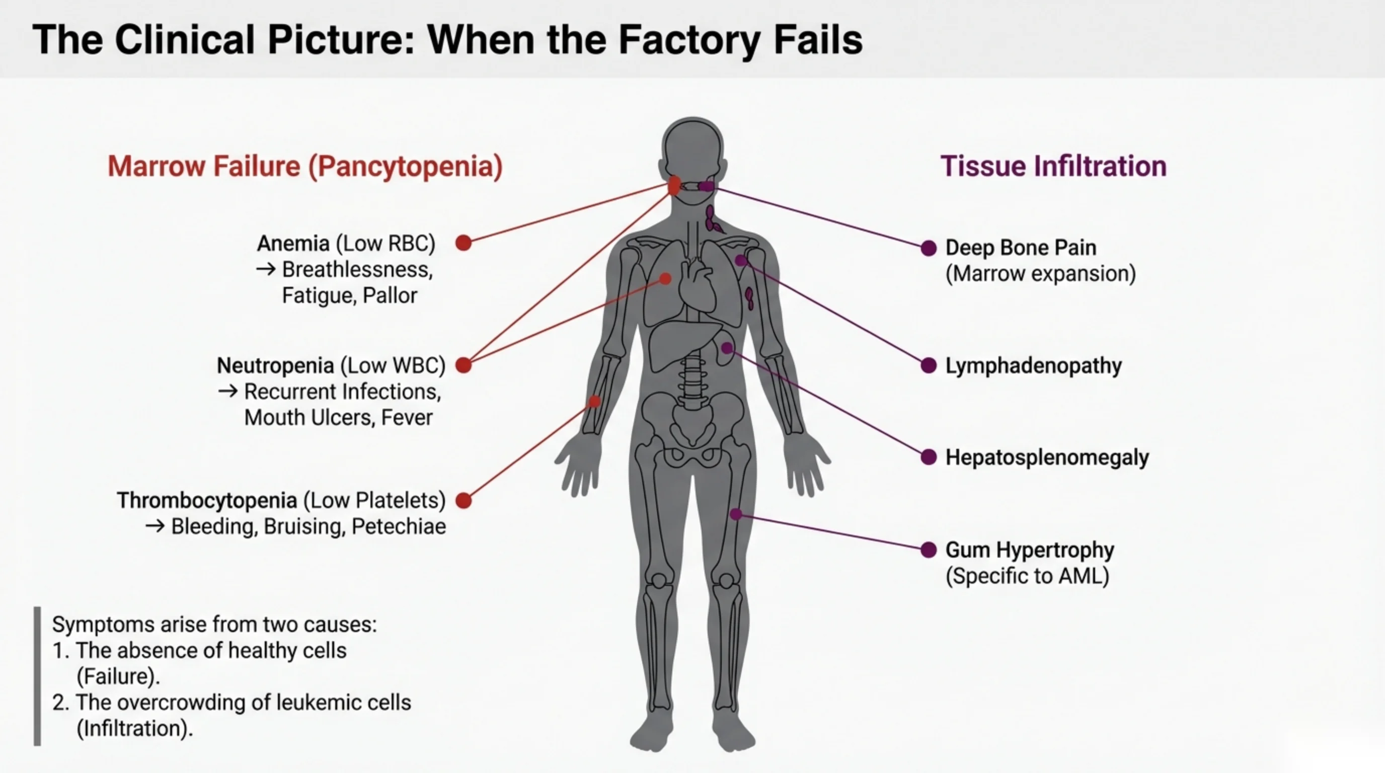
The Clinical Picture: When the Factory Fails
Marrow Failure (Pancytopenia)
- Anemia (Low RBC) → Breathlessness, Fatigue, Pallor
- Neutropenia (Low WBC) → Recurrent Infections, Mouth Ulcers, Fever
- Thrombocytopenia (Low Platelets) → Bleeding, Bruising, Petechiae
Tissue Infiltration
- Deep Bone Pain (Marrow expansion)
- Lymphadenopathy
- Hepatosplenomegaly
- Gum Hypertrophy (Specific to AML)
Symptoms arise from two causes:
- 1. The absence of healthy cells (Failure).
- 2. The overcrowding of leukemic cells (Infiltration).
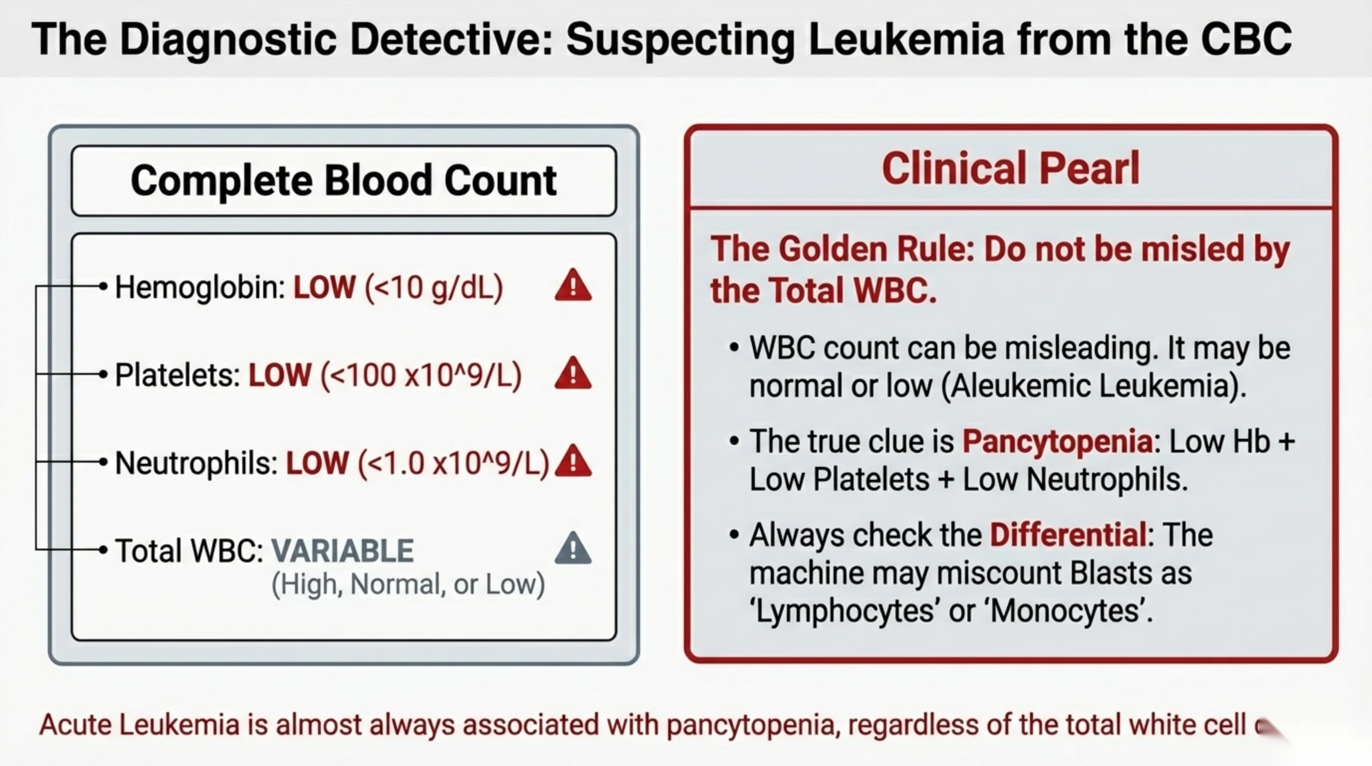
The Diagnostic Detective: Suspecting Leukemia from the CBC
Complete Blood Count
- Hemoglobin: LOW (<10 g/dL)
- Platelets: LOW (<100 x10^9/L)
- Neutrophils: LOW (<1.0 x10^9/L)
- Total WBC: VARIABLE (High, Normal, or Low)
Clinical Pearl — The Golden Rule: Do not be misled by the Total WBC.
- WBC count can be misleading. It may be normal or low (Aleukemic Leukemia).
- The true clue is Pancytopenia: Low Hb + Low Platelets + Low Neutrophils.
- Always check the Differential: The machine may miscount Blasts as ‘Lymphocytes’ or ‘Monocytes’.
Acute Leukemia is almost always associated with pancytopenia, regardless of the total white cell count. (Bottom line partially cut off at slide edge.)
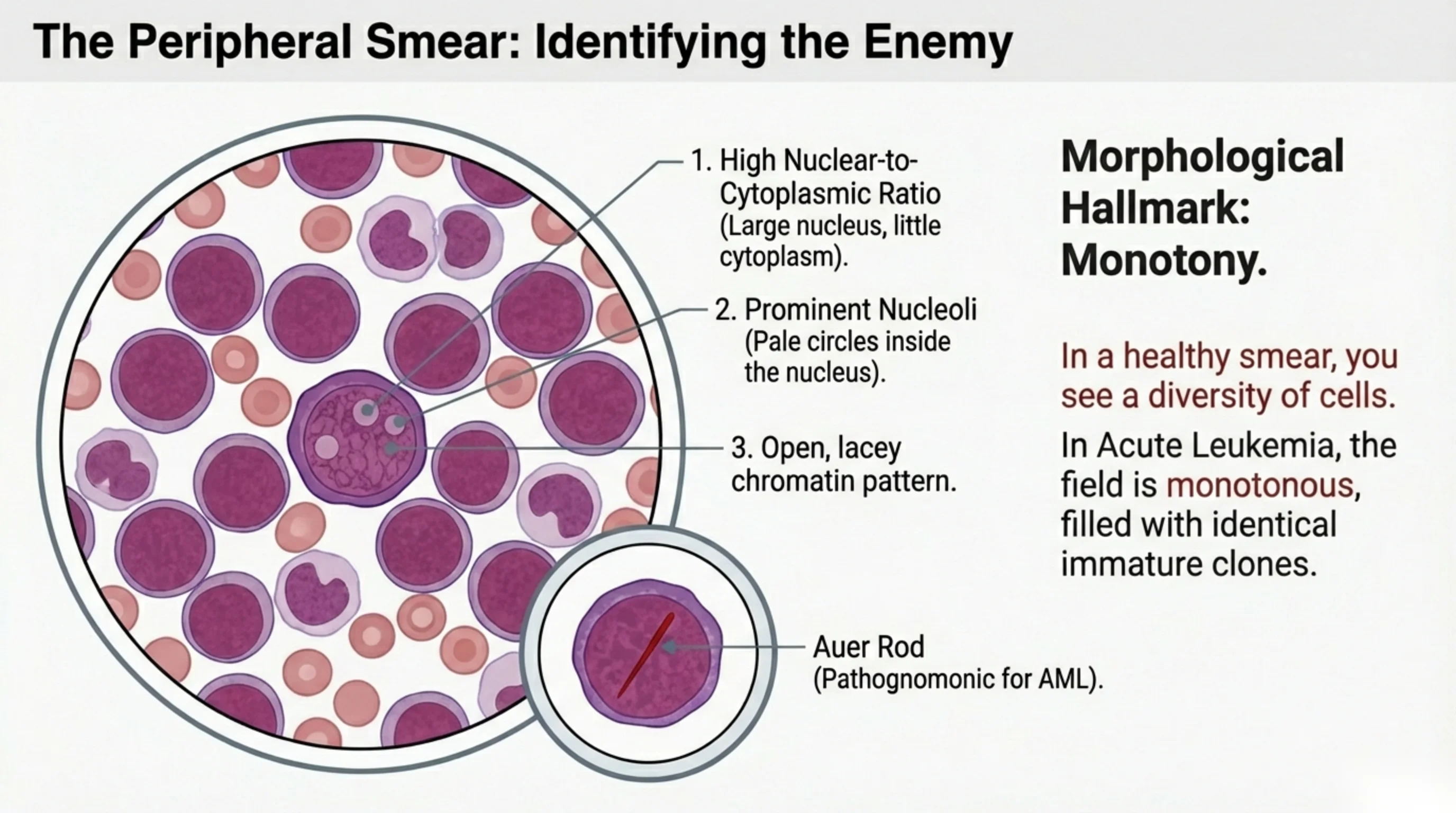
The Peripheral Smear: Identifying the Enemy
Numbered features of the leukemic blast:
- 1. High Nuclear-to-Cytoplasmic Ratio (Large nucleus, little cytoplasm).
- 2. Prominent Nucleoli (Pale circles inside the nucleus).
- 3. Open, lacey chromatin pattern.
Inset label: Auer Rod (Pathognomonic for AML).
Morphological Hallmark: Monotony.
In a healthy smear, you see a diversity of cells.
In Acute Leukemia, the field is monotonous, filled with identical immature clones.
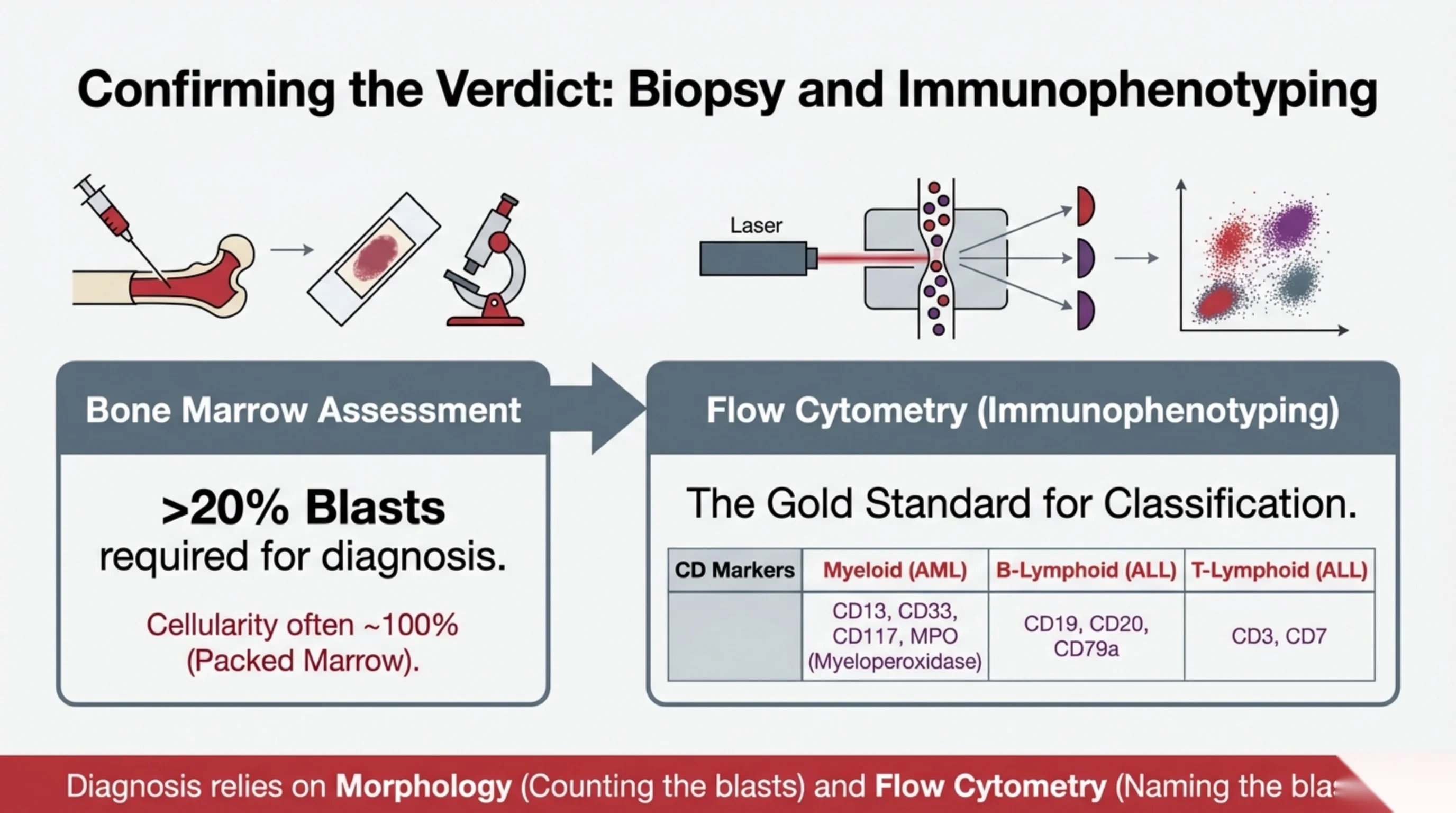
Confirming the Verdict: Biopsy and Immunophenotyping
Bone Marrow Assessment
>20% Blasts required for diagnosis.
Cellularity often ~100% (Packed Marrow).
Flow Cytometry (Immunophenotyping) — The Gold Standard for Classification.
| CD Markers | Myeloid (AML) | B-Lymphoid (ALL) | T-Lymphoid (ALL) |
|---|---|---|---|
| CD13, CD33, CD117, MPO (Myeloperoxidase) | CD19, CD20, CD79a | CD3, CD7 |
Diagnosis relies on Morphology (Counting the blasts) and Flow Cytometry (Naming the blasts). (Bottom line partially cut off at slide edge.)
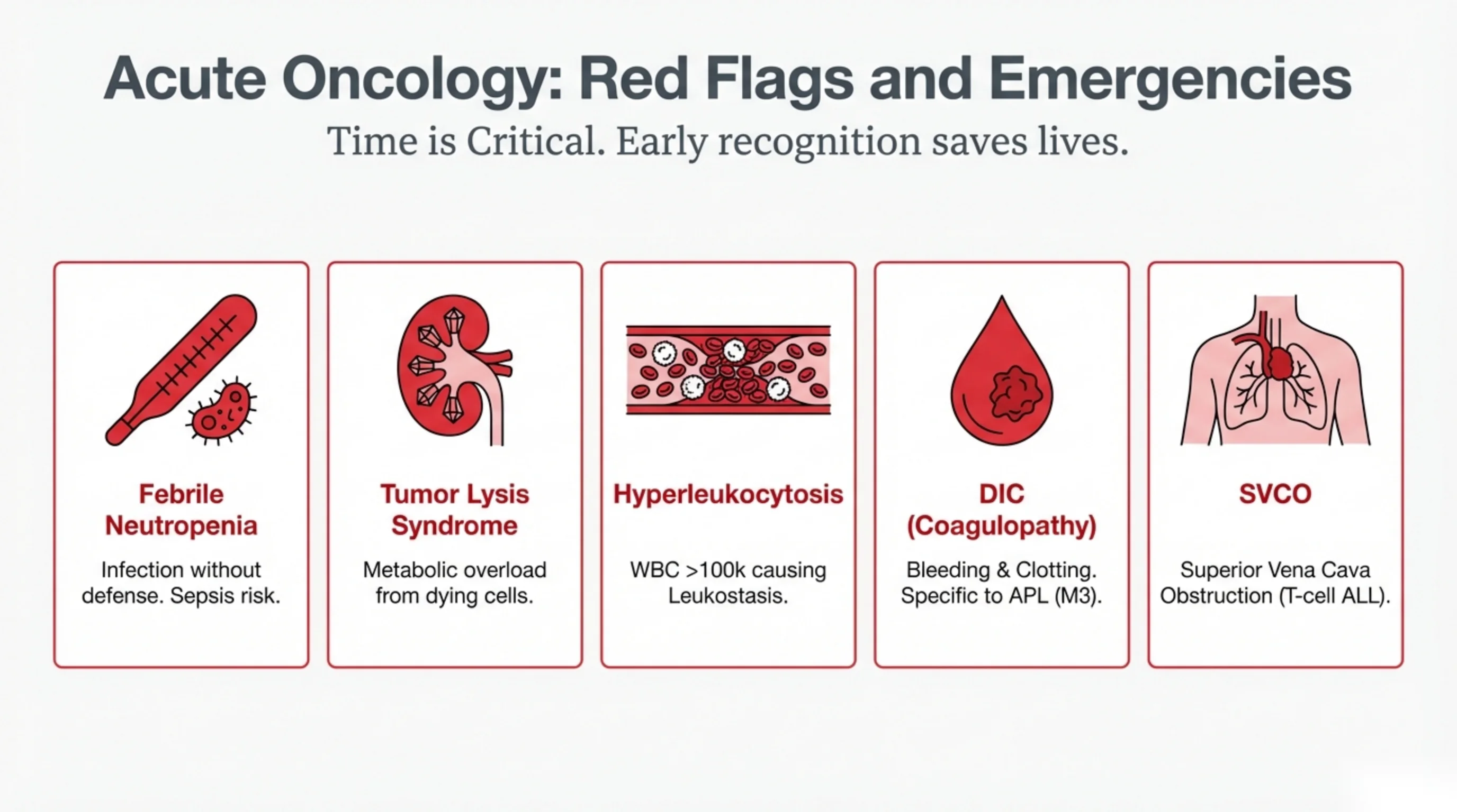
Acute Oncology: Red Flags and Emergencies
Time is Critical. Early recognition saves lives.
- Febrile Neutropenia: Infection without defense. Sepsis risk.
- Tumor Lysis Syndrome: Metabolic overload from dying cells.
- Hyperleukocytosis: WBC >100k causing Leukostasis.
- DIC (Coagulopathy): Bleeding & Clotting. Specific to APL (M3).
- SVCO: Superior Vena Cava Obstruction (T-cell ALL).
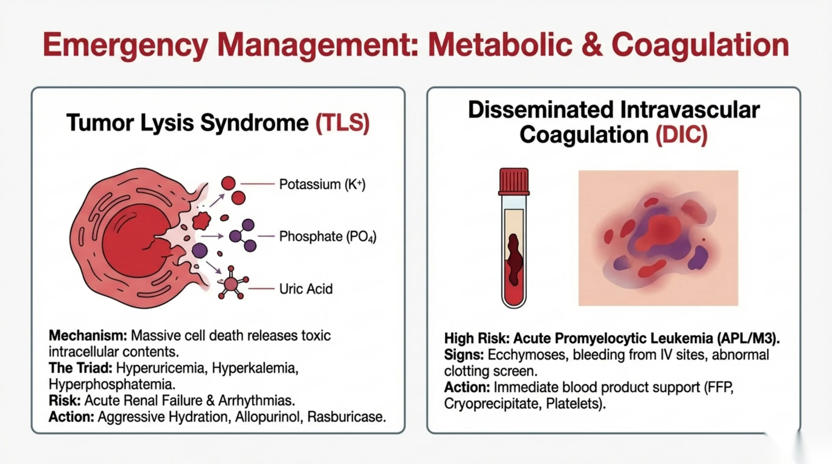
Emergency Management: Metabolic & Coagulation
Tumor Lysis Syndrome (TLS)
The lysing cell releases: Potassium (K+); Phosphate (PO4); Uric Acid.
- Mechanism: Massive cell death releases toxic intracellular contents.
- The Triad: Hyperuricemia, Hyperkalemia, Hyperphosphatemia.
- Risk: Acute Renal Failure & Arrhythmias.
- Action: Aggressive Hydration, Allopurinol, Rasburicase.
Disseminated Intravascular Coagulation (DIC)
- High Risk: Acute Promyelocytic Leukemia (APL/M3).
- Signs: Ecchymoses, bleeding from IV sites, abnormal clotting screen.
- Action: Immediate blood product support (FFP, Cryoprecipitate, Platelets).
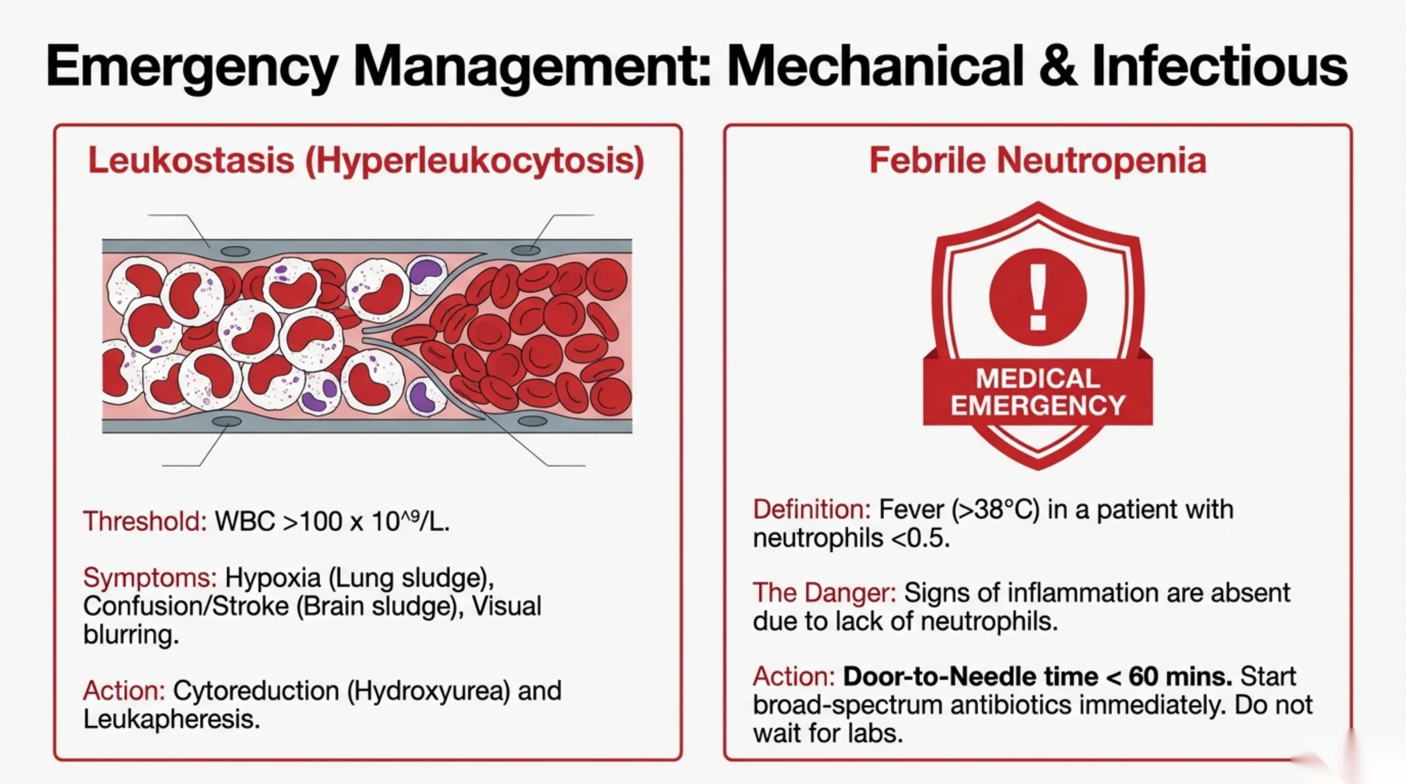
Emergency Management: Mechanical & Infectious
Leukostasis (Hyperleukocytosis)
- Threshold: WBC >100 x 10^9/L.
- Symptoms: Hypoxia (Lung sludge), Confusion/Stroke (Brain sludge), Visual blurring.
- Action: Cytoreduction (Hydroxyurea) and Leukapheresis.
Febrile Neutropenia (marked MEDICAL EMERGENCY)
- Definition: Fever (>38°C) in a patient with neutrophils <0.5.
- The Danger: Signs of inflammation are absent due to lack of neutrophils.
- Action: Door-to-Needle time < 60 mins. Start broad-spectrum antibiotics immediately. Do not wait for labs.
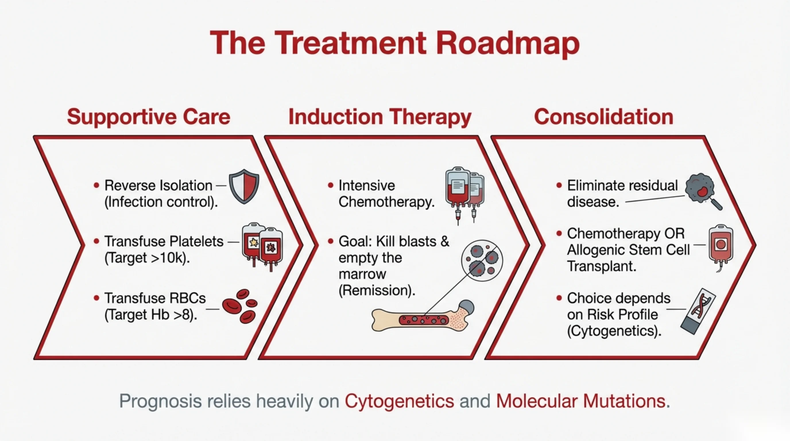
The Treatment Roadmap
Supportive Care
- Reverse Isolation (Infection control).
- Transfuse Platelets (Target >10k).
- Transfuse RBCs (Target Hb >8).
Induction Therapy
- Intensive Chemotherapy.
- Goal: Kill blasts & empty the marrow (Remission).
Consolidation
- Eliminate residual disease.
- Chemotherapy OR Allogenic Stem Cell Transplant.
- Choice depends on Risk Profile (Cytogenetics).
Prognosis relies heavily on Cytogenetics and Molecular Mutations.
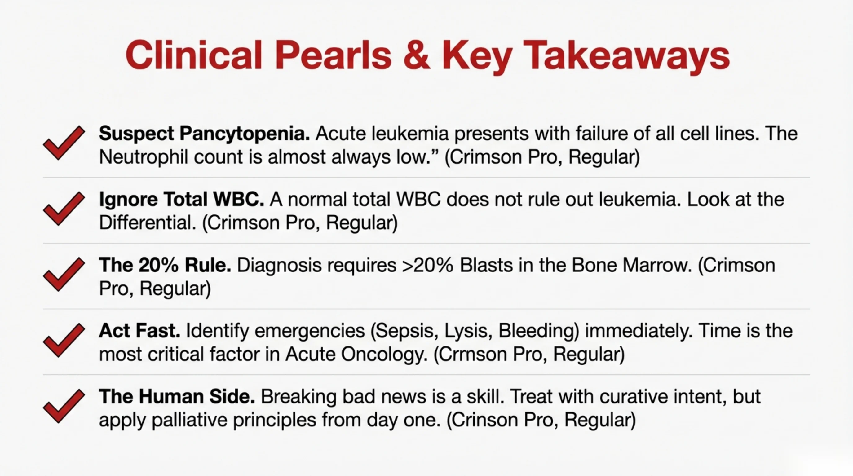
Clinical Pearls & Key Takeaways
- Suspect Pancytopenia. Acute leukemia presents with failure of all cell lines. The Neutrophil count is almost always low.”
- Ignore Total WBC. A normal total WBC does not rule out leukemia. Look at the Differential.
- The 20% Rule. Diagnosis requires >20% Blasts in the Bone Marrow.
- Act Fast. Identify emergencies (Sepsis, Lysis, Bleeding) immediately. Time is the most critical factor in Acute Oncology.
- The Human Side. Breaking bad news is a skill. Treat with curative intent, but apply palliative principles from day one.
(Note: each bullet is followed by a leftover font-placeholder label reading “(Crimson Pro, Regular)”, variously mistyped as “Crmson” and “Crinson”.)
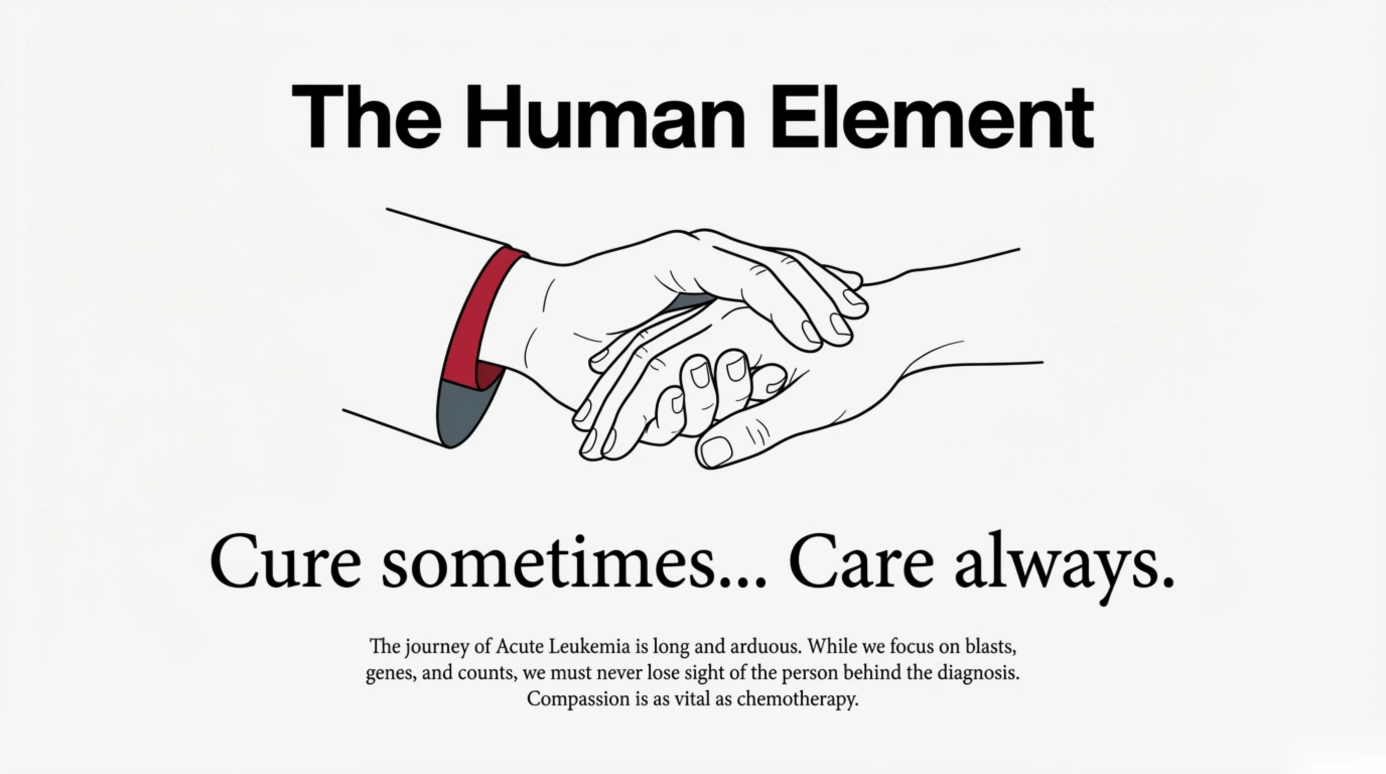
The Human Element
Cure sometimes… Care always.
The journey of Acute Leukemia is long and arduous. While we focus on blasts, genes, and counts, we must never lose sight of the person behind the diagnosis. Compassion is as vital as chemotherapy.