Lymphoma
From pathogenesis to management — classification, presentation, staging, and immunotherapy.
Lymphoma
Malignant Lymphoproliferative Disorders: From Pathogenesis to Clinical Management
Physiology | Classification | Diagnosis | Immunotherapy
Clinical editorial
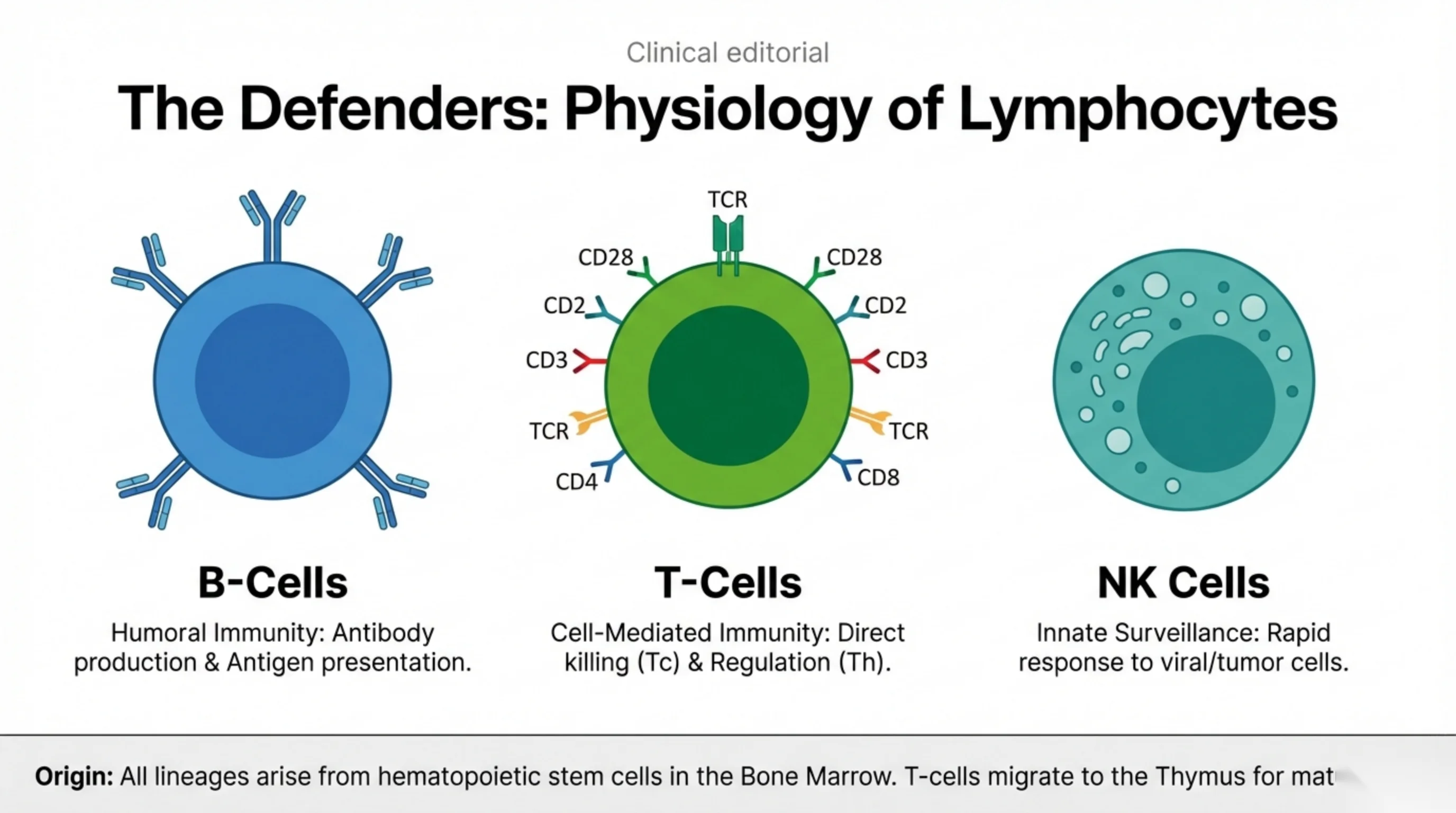
The Defenders: Physiology of Lymphocytes
B-Cells
Humoral Immunity: Antibody production & Antigen presentation.
T-Cells
Surface markers shown on the T-cell diagram: TCR, CD28, CD2, CD3, TCR, CD4, CD8.
Cell-Mediated Immunity: Direct killing (Tc) & Regulation (Th).
NK Cells
Innate Surveillance: Rapid response to viral/tumor cells.
Origin: All lineages arise from hematopoietic stem cells in the Bone Marrow. T-cells migrate to the Thymus for mat… [text truncated at slide edge, presumably "maturation"]
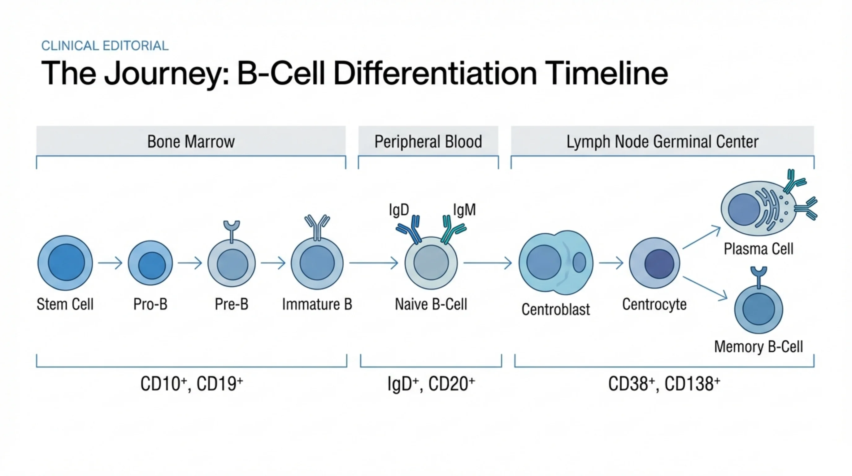
The Journey: B-Cell Differentiation Timeline
Bone Marrow: Stem Cell → Pro-B → Pre-B → Immature B (markers: CD10+, CD19+)
Peripheral Blood: Naive B-Cell, expressing IgD and IgM (markers: IgD+, CD20+)
Lymph Node Germinal Center: Centroblast → Centrocyte → which differentiates into either Plasma Cell or Memory B-Cell (markers: CD38+, CD138+)
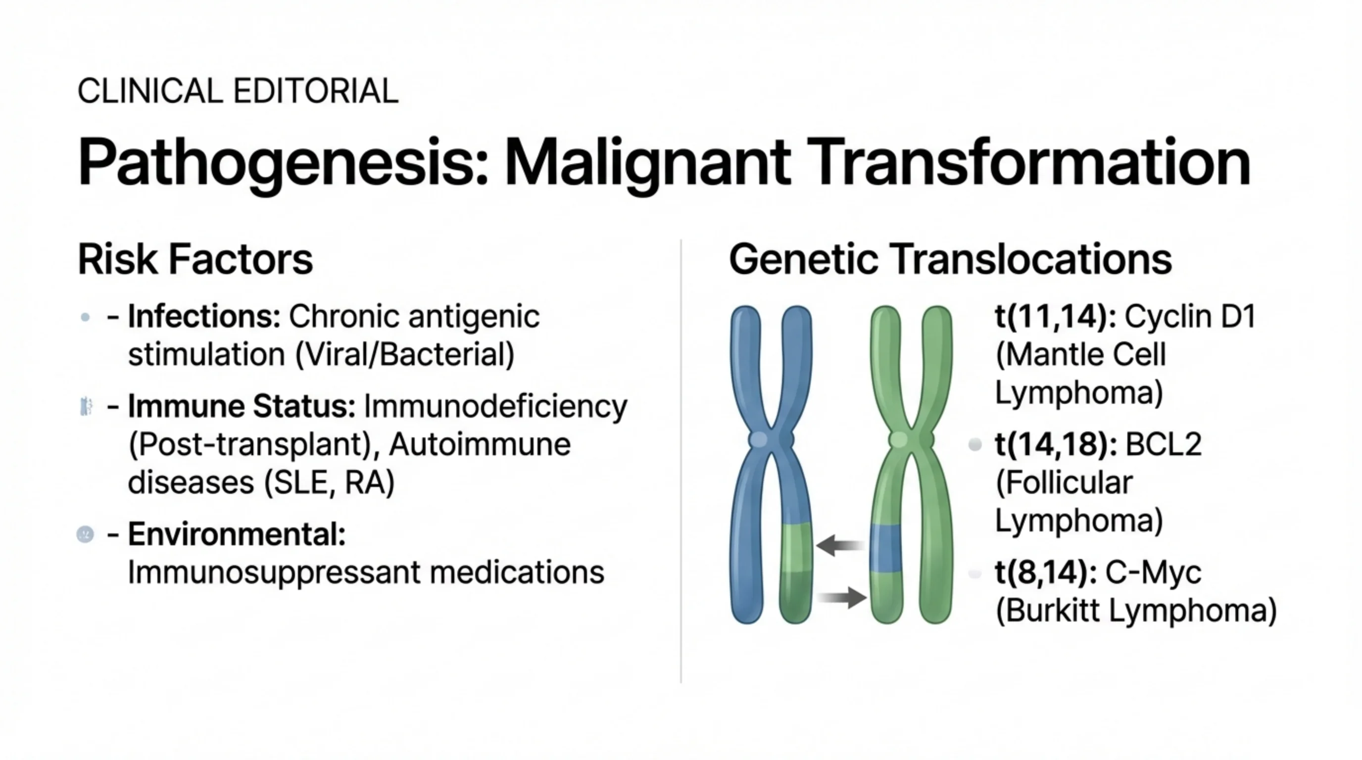
Pathogenesis: Malignant Transformation
Risk Factors
- Infections: Chronic antigenic stimulation (Viral/Bacterial)
- Immune Status: Immunodeficiency (Post-transplant), Autoimmune diseases (SLE, RA)
- Environmental: Immunosuppressant medications
Genetic Translocations
- t(11,14): Cyclin D1 (Mantle Cell Lymphoma)
- t(14,18): BCL2 (Follicular Lymphoma)
- t(8,14): C-Myc (Burkitt Lymphoma)
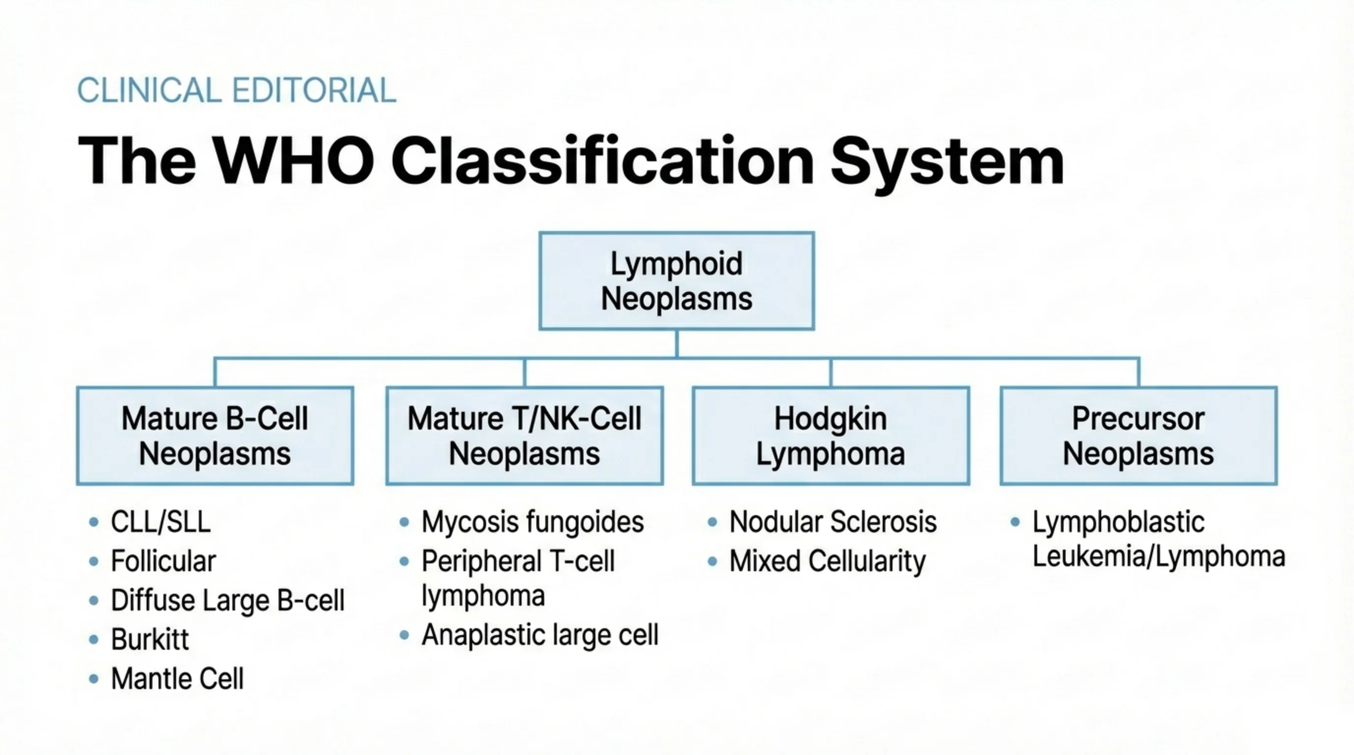
The WHO Classification System
Lymphoid Neoplasms branch into four categories:
Mature B-Cell Neoplasms
- CLL/SLL
- Follicular
- Diffuse Large B-cell
- Burkitt
- Mantle Cell
Mature T/NK-Cell Neoplasms
- Mycosis fungoides
- Peripheral T-cell lymphoma
- Anaplastic large cell
Hodgkin Lymphoma
- Nodular Sclerosis
- Mixed Cellularity
Precursor Neoplasms
- Lymphoblastic Leukemia/Lymphoma
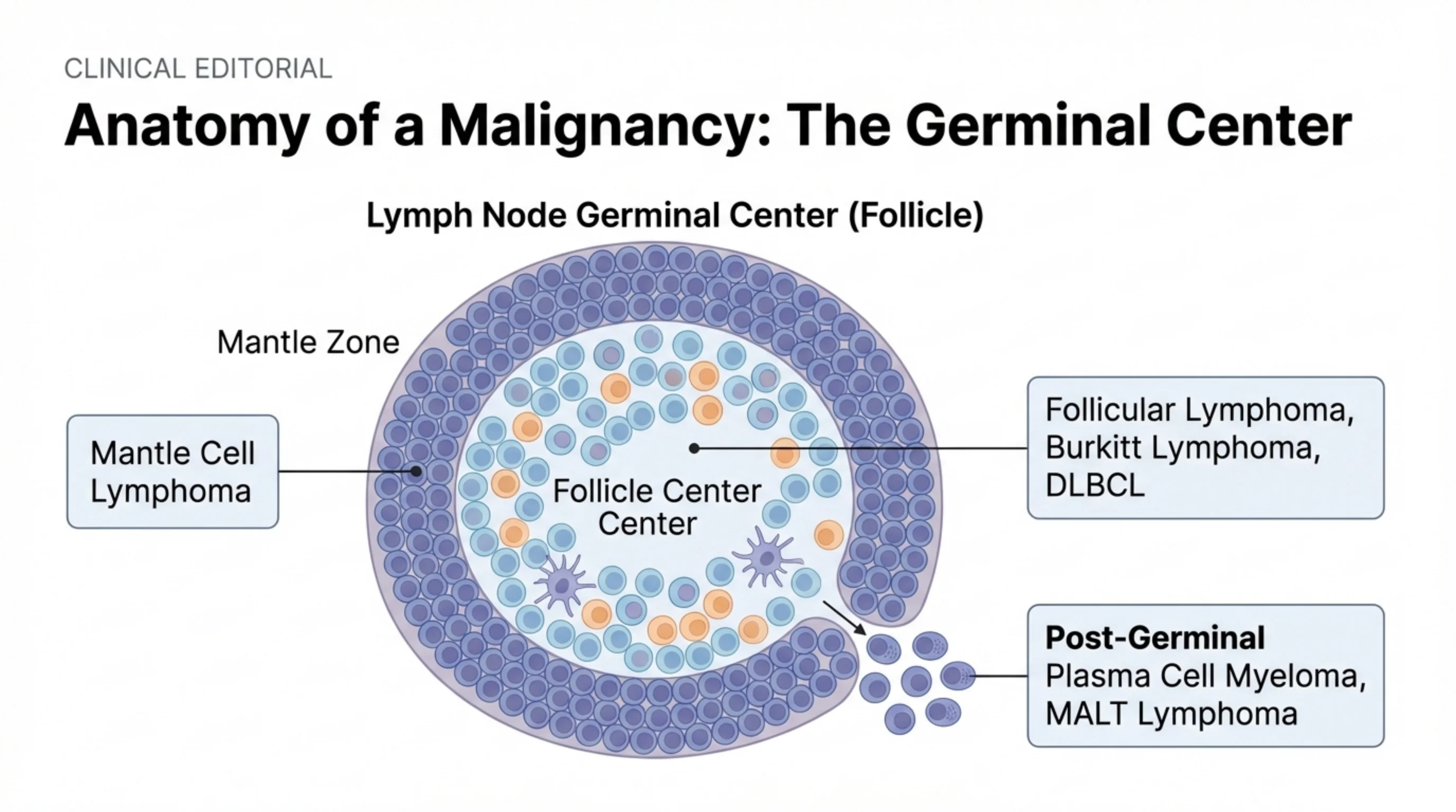
Anatomy of a Malignancy: The Germinal Center
Diagram: Lymph Node Germinal Center (Follicle), with labeled Mantle Zone and Follicle Center Center.
- Mantle Zone → Mantle Cell Lymphoma
- Follicle Center → Follicular Lymphoma, Burkitt Lymphoma, DLBCL
- Post-Germinal (cells exiting the follicle) → Plasma Cell Myeloma, MALT Lymphoma
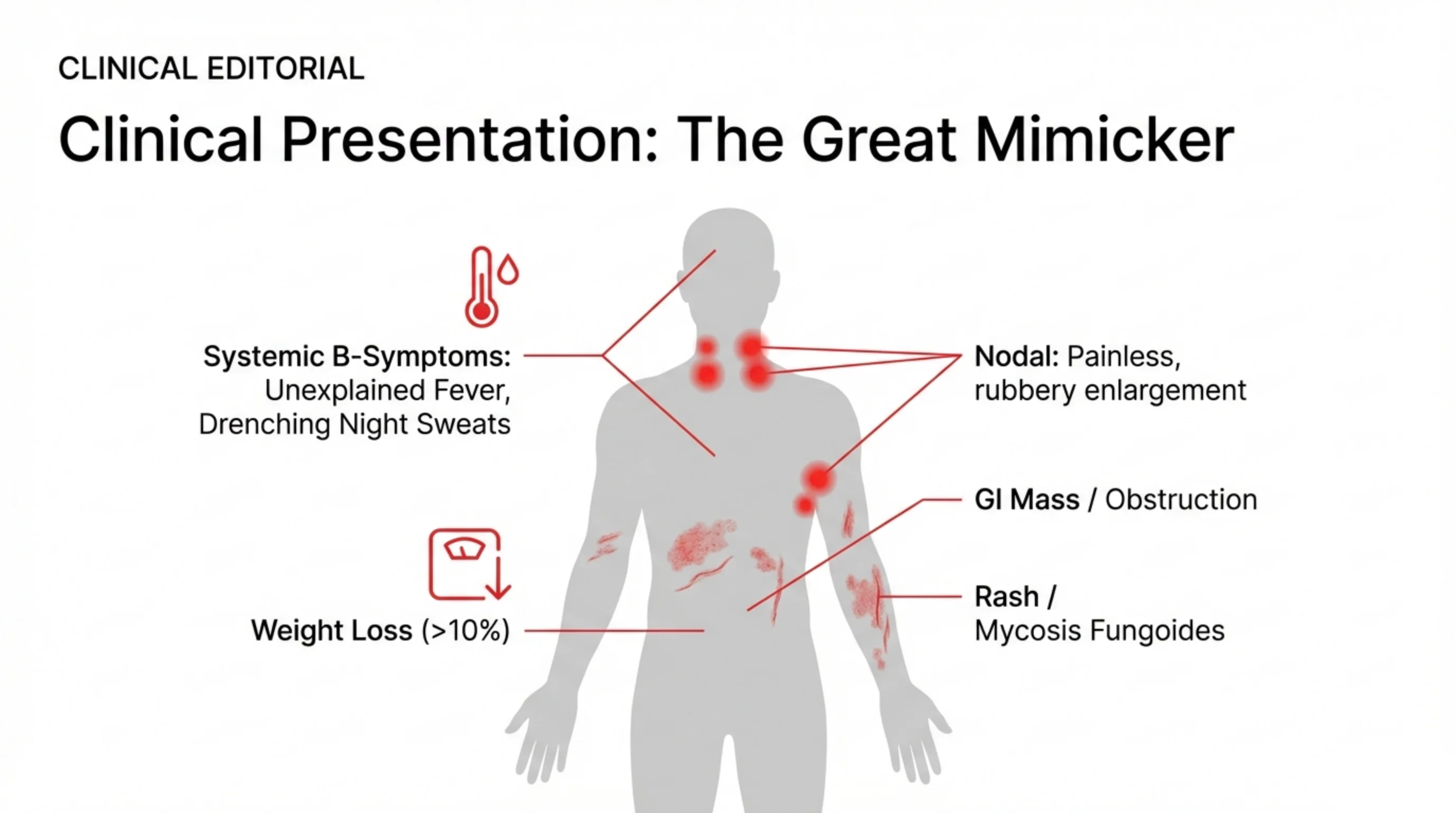
Clinical Presentation: The Great Mimicker
- Systemic B-Symptoms: Unexplained Fever, Drenching Night Sweats
- Weight Loss (>10%)
- Nodal: Painless, rubbery enlargement
- GI Mass / Obstruction
- Rash / Mycosis Fungoides
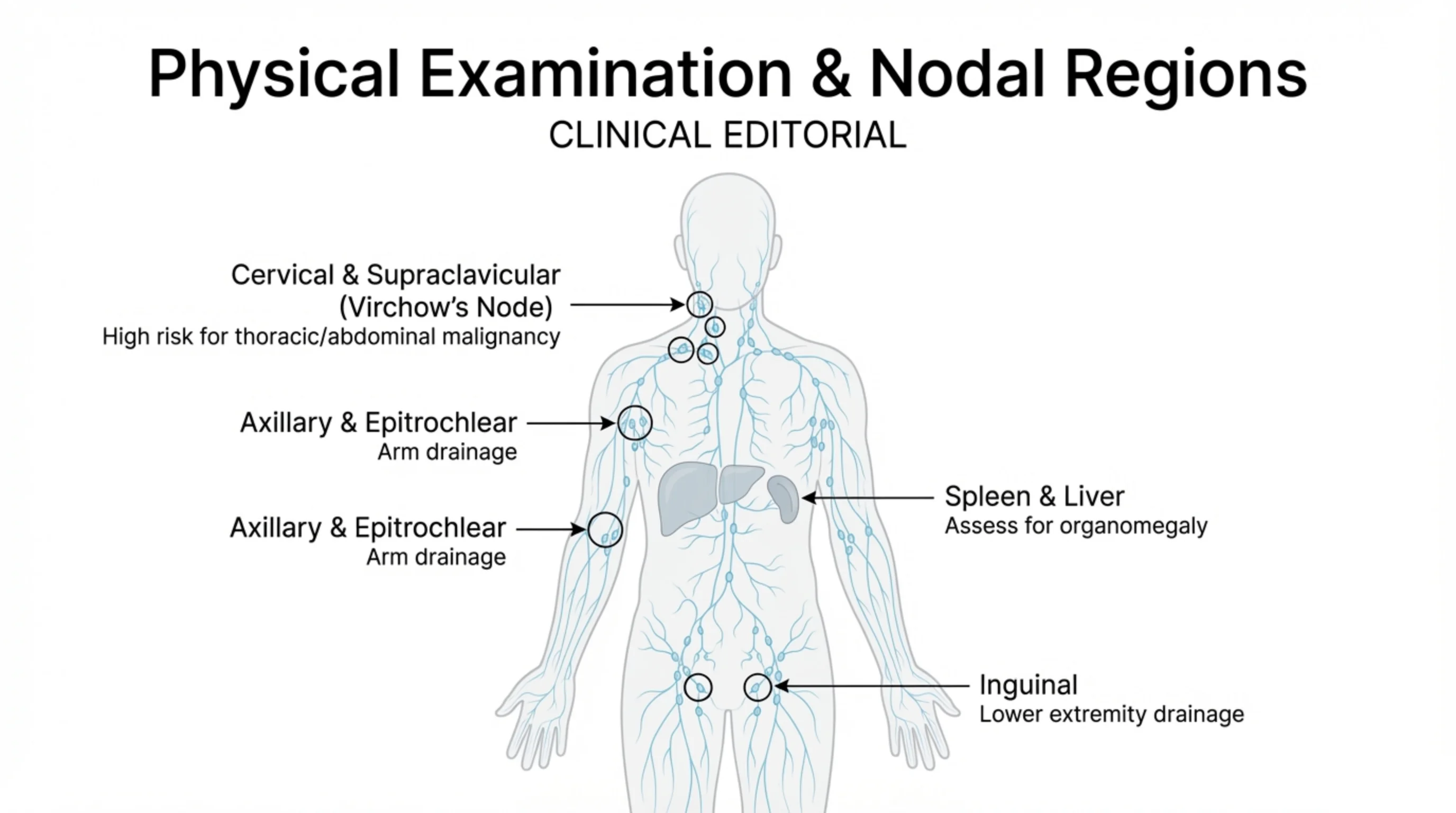
Physical Examination & Nodal Regions
- Cervical & Supraclavicular (Virchow's Node): High risk for thoracic/abdominal malignancy
- Axillary & Epitrochlear: Arm drainage
- Axillary & Epitrochlear: Arm drainage
- Spleen & Liver: Assess for organomegaly
- Inguinal: Lower extremity drainage
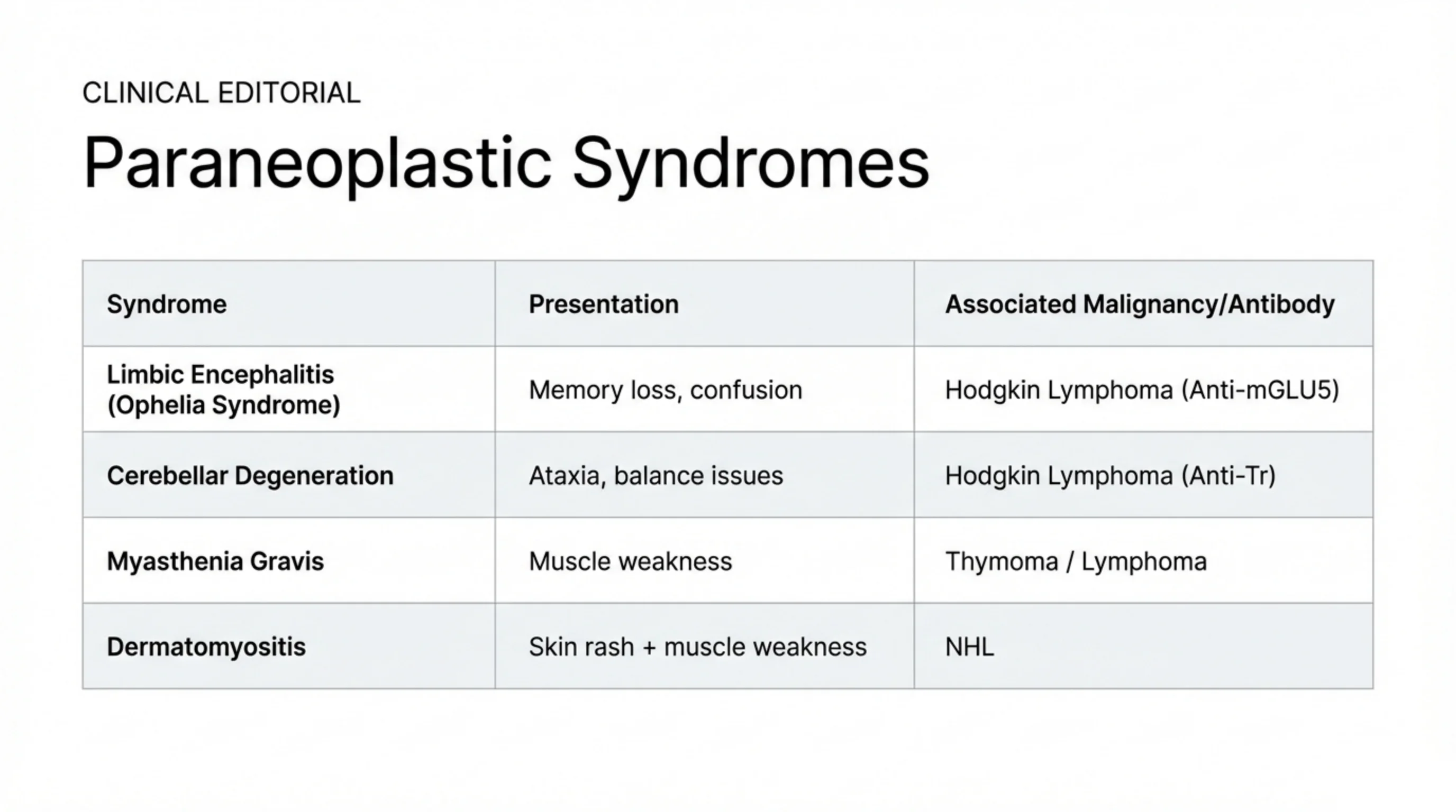
Paraneoplastic Syndromes
| Syndrome | Presentation | Associated Malignancy/Antibody |
|---|---|---|
| Limbic Encephalitis (Ophelia Syndrome) | Memory loss, confusion | Hodgkin Lymphoma (Anti-mGLU5) |
| Cerebellar Degeneration | Ataxia, balance issues | Hodgkin Lymphoma (Anti-Tr) |
| Myasthenia Gravis | Muscle weakness | Thymoma / Lymphoma |
| Dermatomyositis | Skin rash + muscle weakness | NHL |
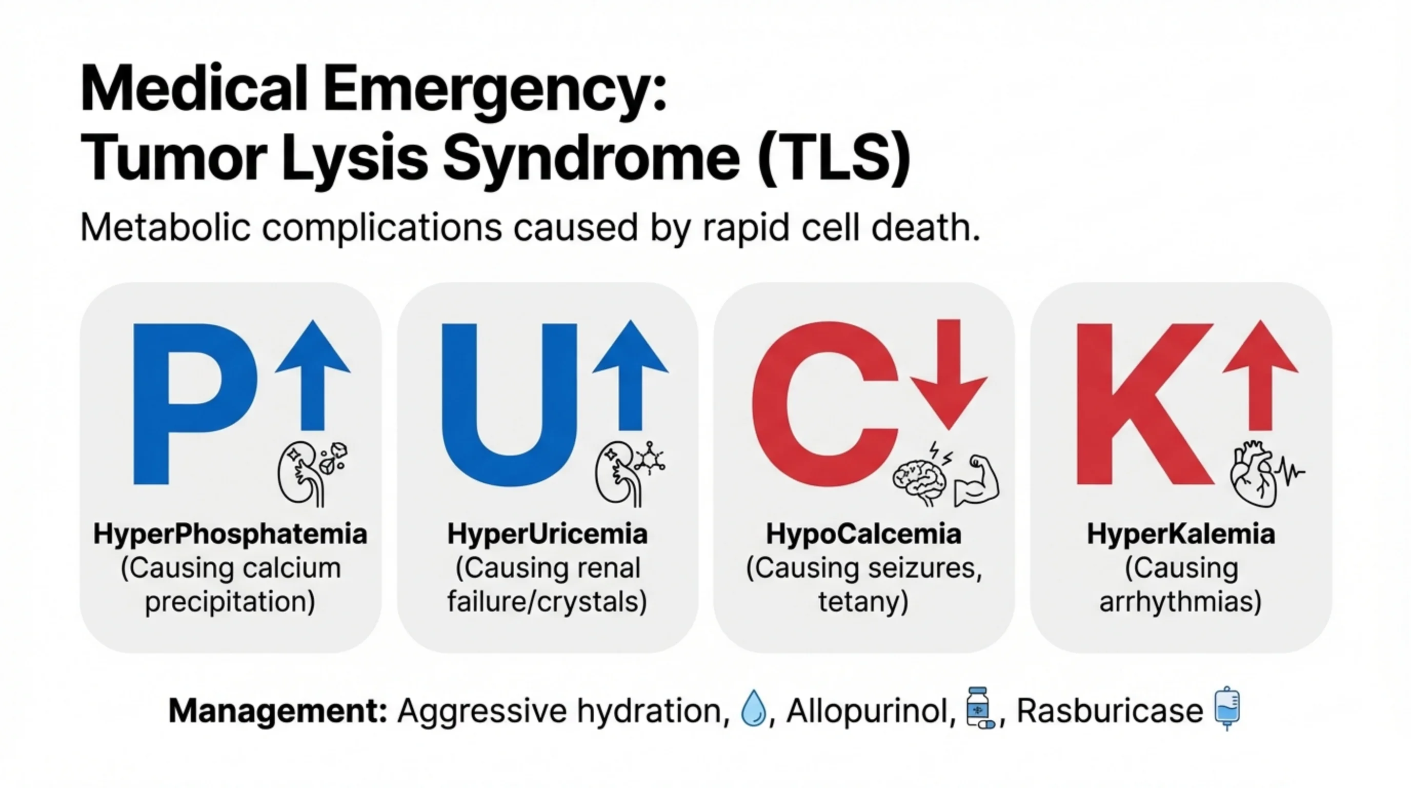
Medical Emergency: Tumor Lysis Syndrome (TLS)
Metabolic complications caused by rapid cell death.
- P – HyperPhosphatemia (increased): Causing calcium precipitation
- U – HyperUricemia (increased): Causing renal failure/crystals
- C – HypoCalcemia (decreased): Causing seizures, tetany
- K – HyperKalemia (increased): Causing arrhythmias
Management: Aggressive hydration, Allopurinol, Rasburicase
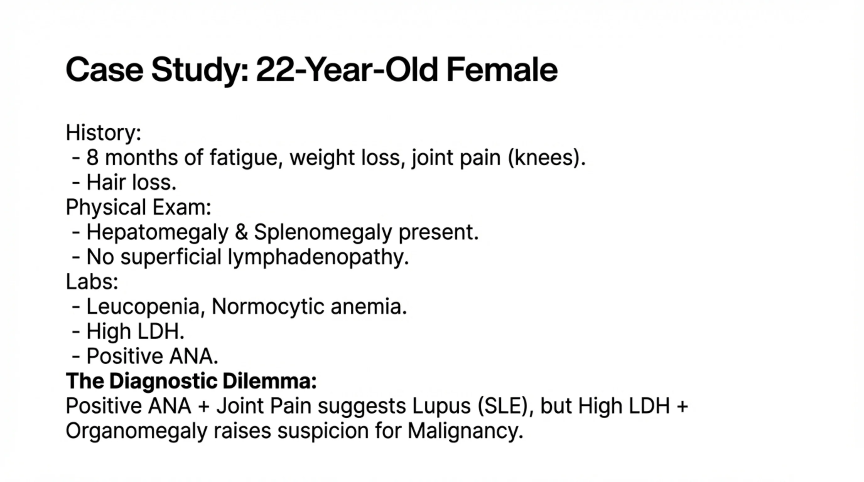
Case Study: 22-Year-Old Female
History:
- 8 months of fatigue, weight loss, joint pain (knees).
- Hair loss.
Physical Exam:
- Hepatomegaly & Splenomegaly present.
- No superficial lymphadenopathy.
Labs:
- Leucopenia, Normocytic anemia.
- High LDH.
- Positive ANA.
The Diagnostic Dilemma: Positive ANA + Joint Pain suggests Lupus (SLE), but High LDH + Organomegaly raises suspicion for Malignancy.
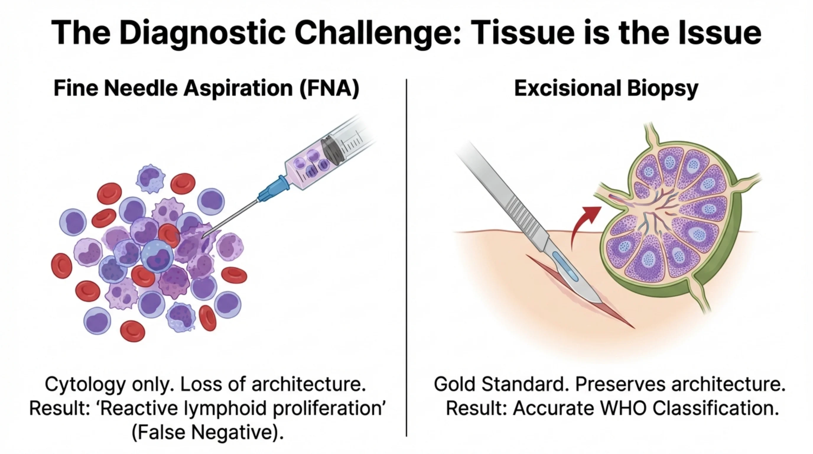
The Diagnostic Challenge: Tissue is the Issue
Fine Needle Aspiration (FNA)
Cytology only. Loss of architecture. Result: 'Reactive lymphoid proliferation' (False Negative).
Excisional Biopsy
Gold Standard. Preserves architecture. Result: Accurate WHO Classification.
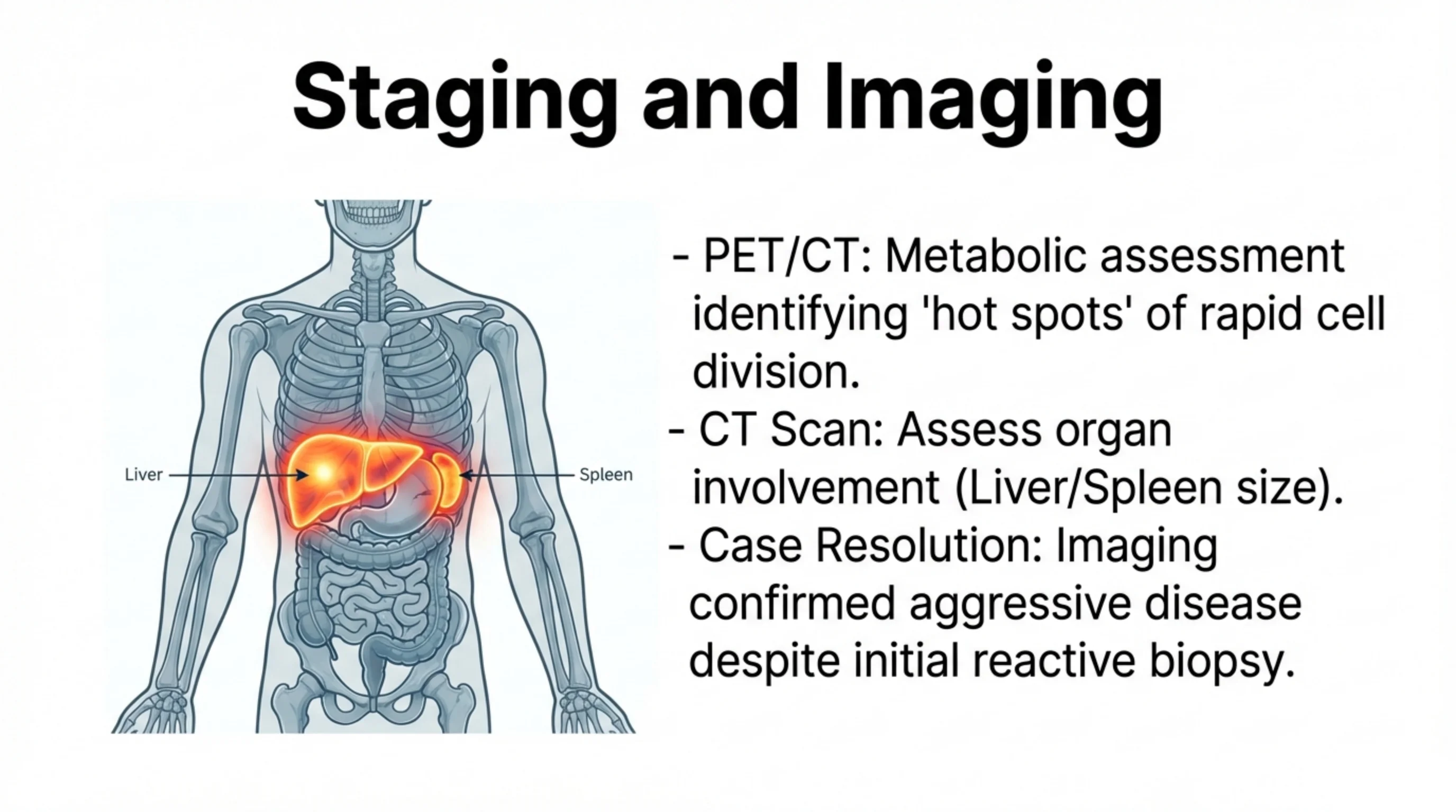
Staging and Imaging
Illustration labels: Liver and Spleen.
- PET/CT: Metabolic assessment identifying 'hot spots' of rapid cell division.
- CT Scan: Assess organ involvement (Liver/Spleen size).
- Case Resolution: Imaging confirmed aggressive disease despite initial reactive biopsy.
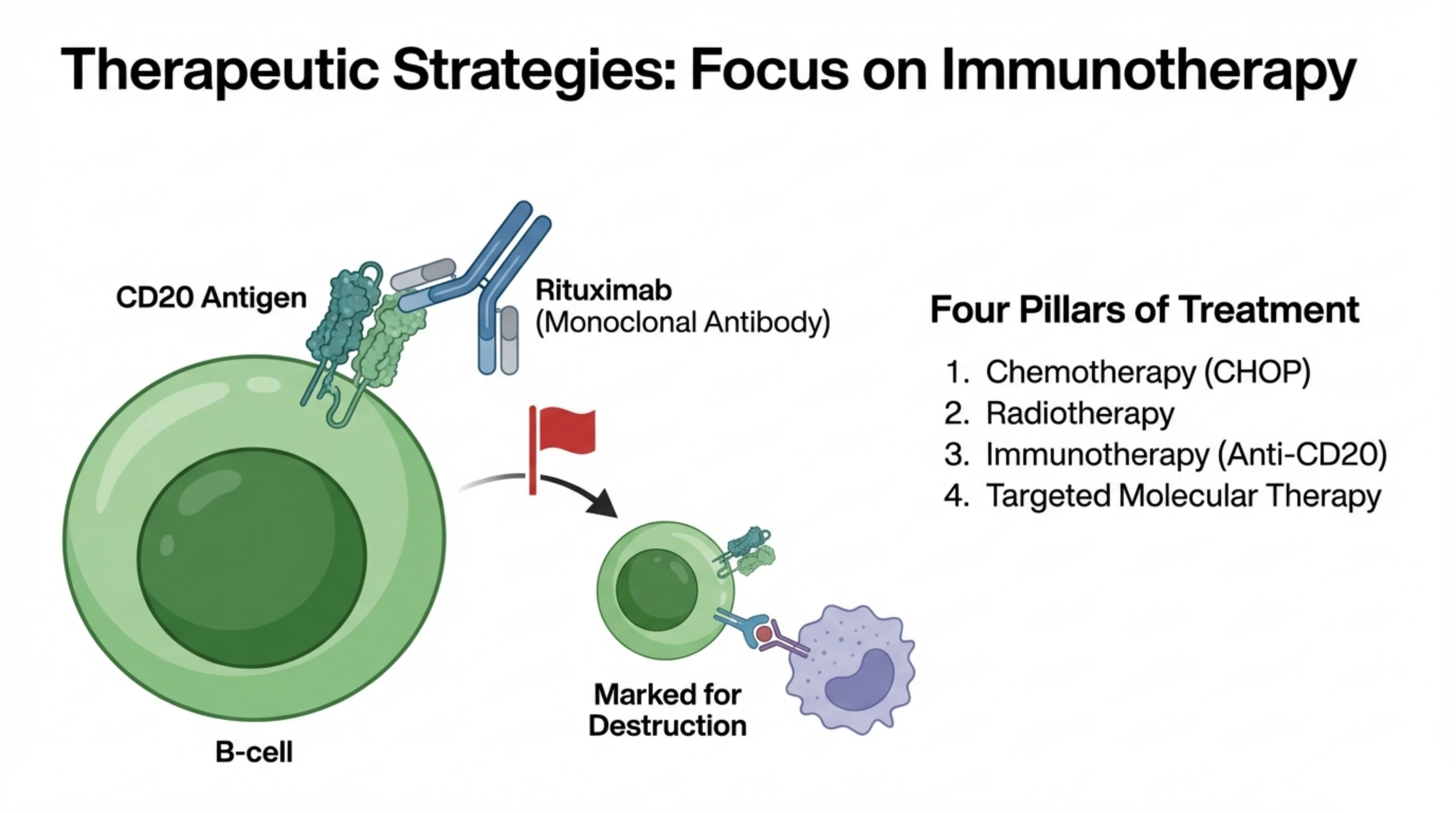
Therapeutic Strategies: Focus on Immunotherapy
Diagram: Rituximab (Monoclonal Antibody) binds the CD20 Antigen on a B-cell, which is then Marked for Destruction.
Four Pillars of Treatment
- Chemotherapy (CHOP)
- Radiotherapy
- Immunotherapy (Anti-CD20)
- Targeted Molecular Therapy
Clinical Takeaways
- Definition: Malignancy of mature lymphocytes arising in lymph nodes or extranodal tissue.
- Pathogenesis: Driven by genetic translocations (e.g., t(14,18)) and immune dysregulation.
- Diagnosis: The "Great Mimicker." Excisional biopsy is mandatory to assess nodal architecture.
- Emergency: Monitor for Tumor Lysis Syndrome (Hyperkalemia, Hyperuricemia).
- Treatment: Accurate histological classification guides targeted immunotherapy (Anti-CD20).