Myeloid Neoplasms
Myeloproliferative neoplasms and myelodysplastic syndromes — classification and management.

Myeloproliferative Neoplasms (MPNs) & Myelodysplastic Syndromes (MDS)
A Clinical Guide to Classification, Pathophysiology, and Management
“For the learners… Classify.”
Based on Student Notes / HK 2022
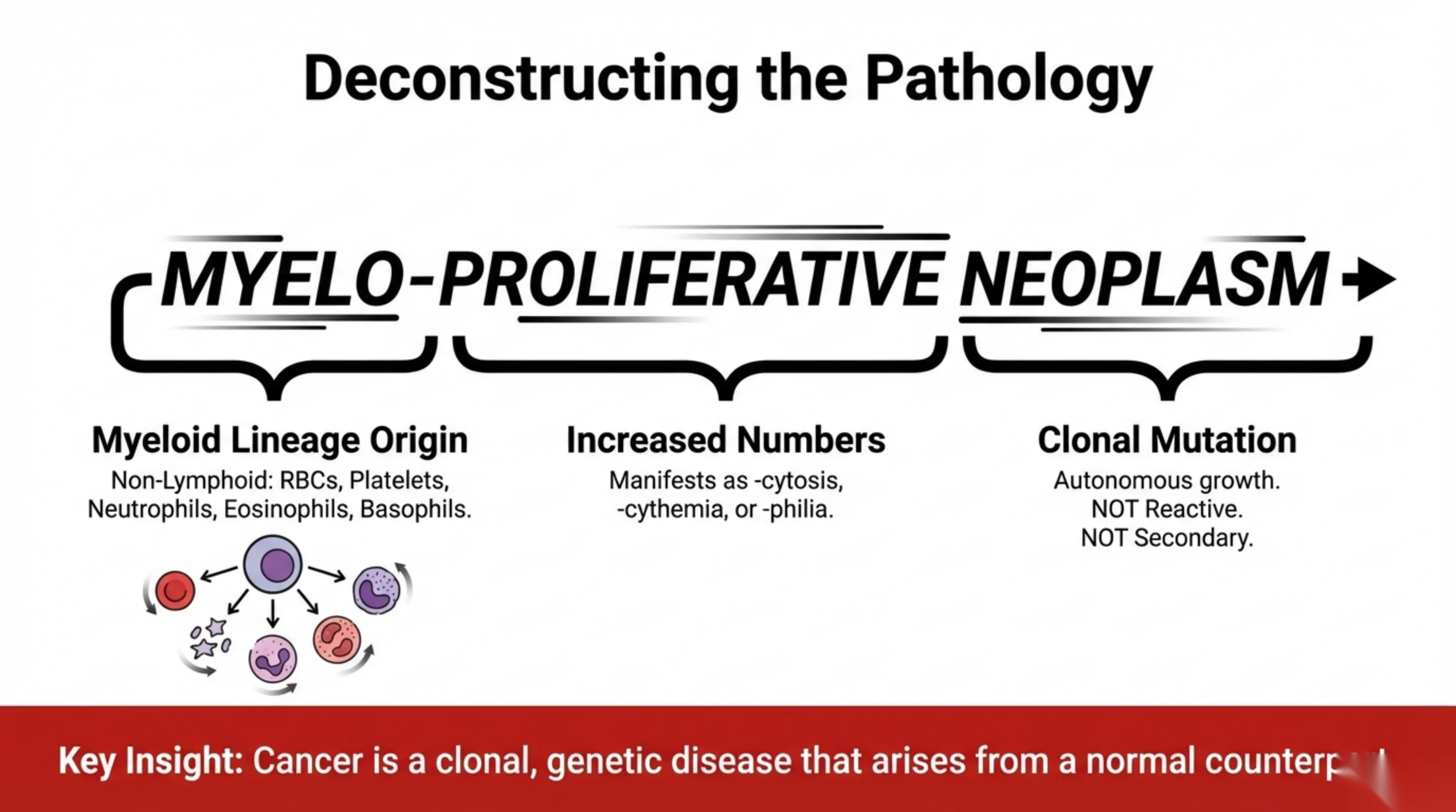
Deconstructing the Pathology
The term MYELO-PROLIFERATIVE NEOPLASM is deconstructed into three components:
- Myeloid Lineage Origin — Non-Lymphoid: RBCs, Platelets, Neutrophils, Eosinophils, Basophils.
- Increased Numbers — Manifests as -cytosis, -cythemia, or -philia.
- Clonal Mutation — Autonomous growth. NOT Reactive. NOT Secondary.
Key Insight: Cancer is a clonal, genetic disease that arises from a normal counterp[art] [text cut off at slide edge].
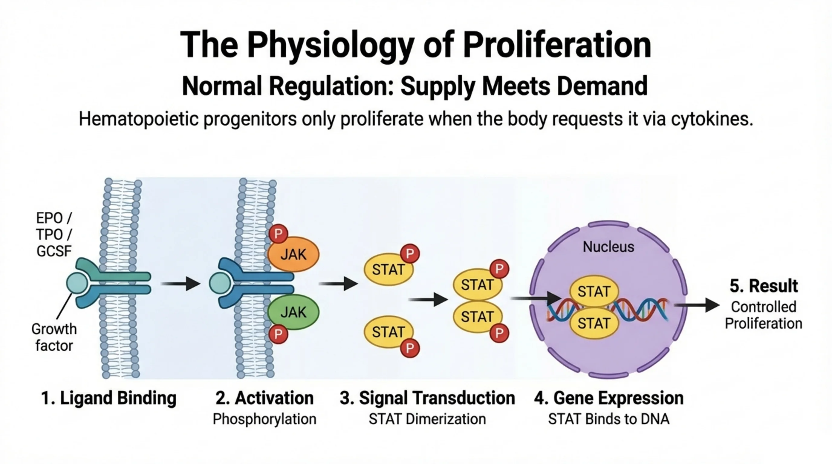
The Physiology of Proliferation
Normal Regulation: Supply Meets Demand
Hematopoietic progenitors only proliferate when the body requests it via cytokines.
Signaling cascade (growth factors EPO / TPO / GCSF):
- Ligand Binding — growth factor binds the receptor.
- Activation — Phosphorylation (JAK).
- Signal Transduction — STAT Dimerization.
- Gene Expression — STAT Binds to DNA (in the Nucleus).
- Result — Controlled Proliferation.
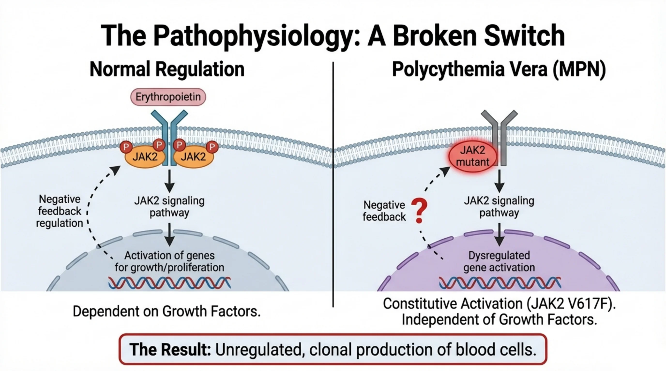
The Pathophysiology: A Broken Switch
Normal Regulation
- Erythropoietin binds its receptor; phosphorylated JAK2 dimers drive the JAK2 signaling pathway.
- Leads to Activation of genes for growth/proliferation.
- Negative feedback regulation is intact.
- Dependent on Growth Factors.
Polycythemia Vera (MPN)
- JAK2 mutant drives the JAK2 signaling pathway without ligand.
- Leads to Dysregulated gene activation.
- Negative feedback is lost (indicated by “?”).
- Constitutive Activation (JAK2 V617F). Independent of Growth Factors.
The Result: Unregulated, clonal production of blood cells.
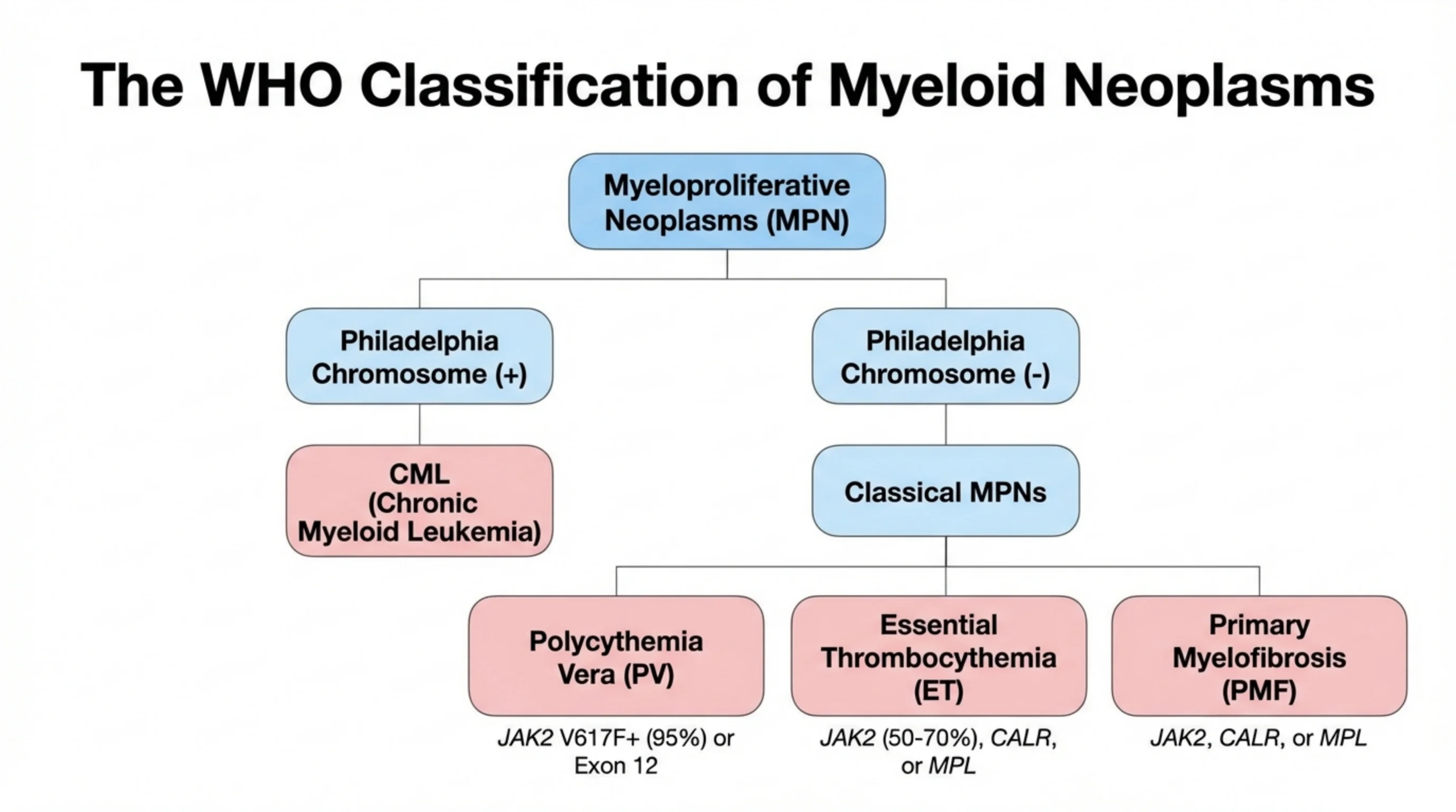
The WHO Classification of Myeloid Neoplasms
Myeloproliferative Neoplasms (MPN) divide by Philadelphia chromosome status:
- Philadelphia Chromosome (+)
- CML (Chronic Myeloid Leukemia)
- Philadelphia Chromosome (−) → Classical MPNs:
- Polycythemia Vera (PV) — JAK2 V617F+ (95%) or Exon 12
- Essential Thrombocythemia (ET) — JAK2 (50-70%), CALR, or MPL
- Primary Myelofibrosis (PMF) — JAK2, CALR, or MPL
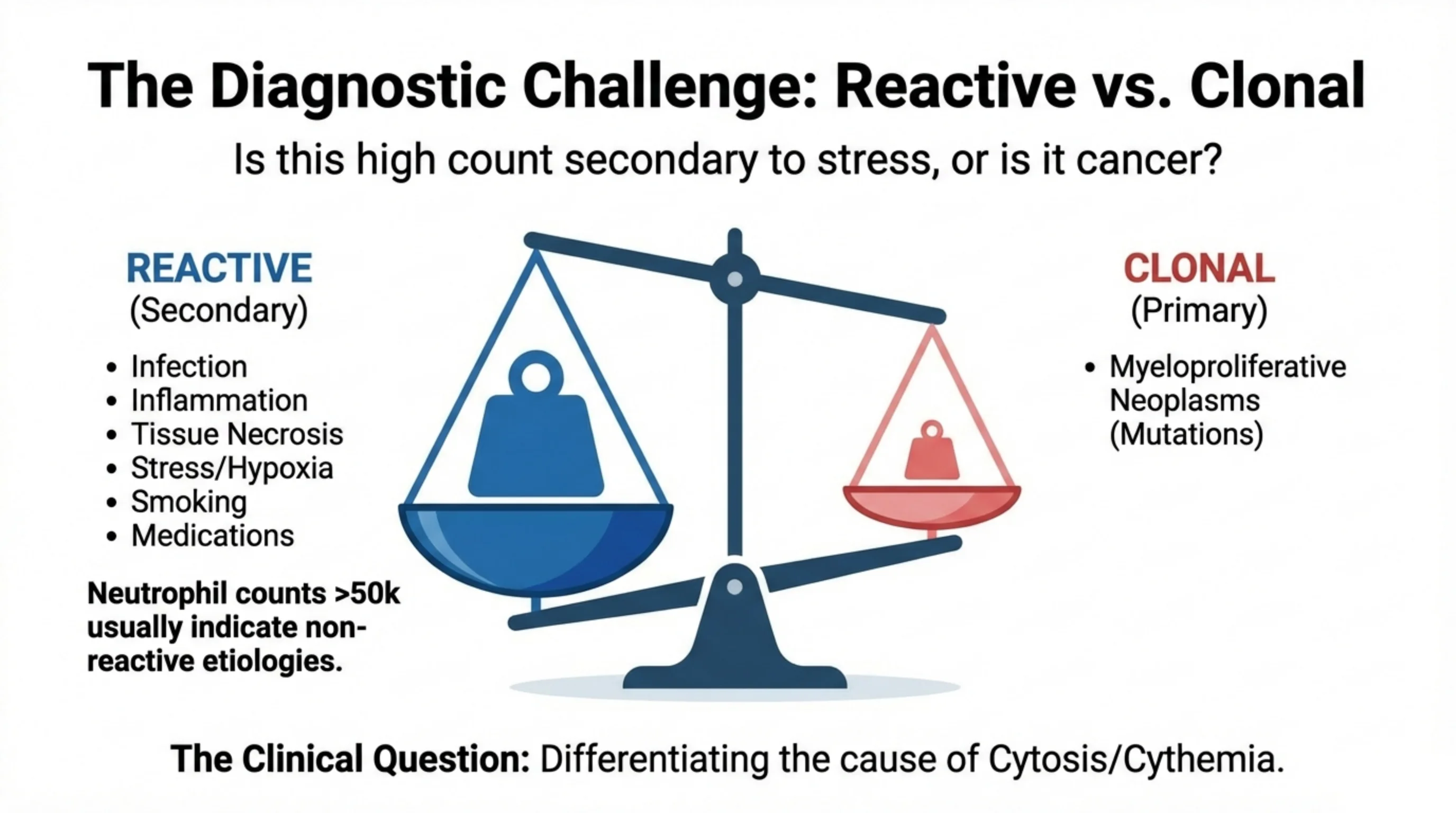
The Diagnostic Challenge: Reactive vs. Clonal
Is this high count secondary to stress, or is it cancer?
REACTIVE (Secondary)
- Infection
- Inflammation
- Tissue Necrosis
- Stress/Hypoxia
- Smoking
- Medications
Neutrophil counts >50k usually indicate non-reactive etiologies.
CLONAL (Primary)
- Myeloproliferative Neoplasms (Mutations)
The Clinical Question: Differentiating the cause of Cytosis/Cythemia.
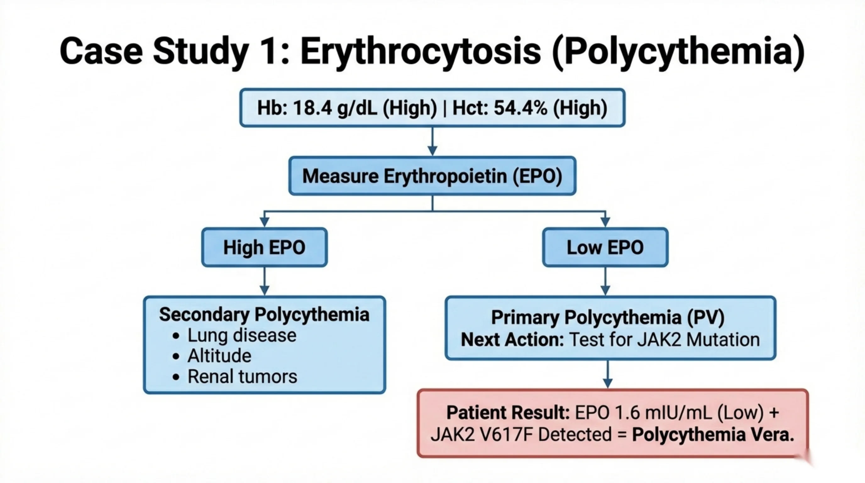
Case Study 1: Erythrocytosis (Polycythemia)
Hb: 18.4 g/dL (High) | Hct: 54.4% (High)
Next step: Measure Erythropoietin (EPO).
- High EPO → Secondary Polycythemia:
- Lung disease
- Altitude
- Renal tumors
- Low EPO → Primary Polycythemia (PV). Next Action: Test for JAK2 Mutation.
Patient Result: EPO 1.6 mIU/mL (Low) + JAK2 V617F Detected = Polycythemia Vera.
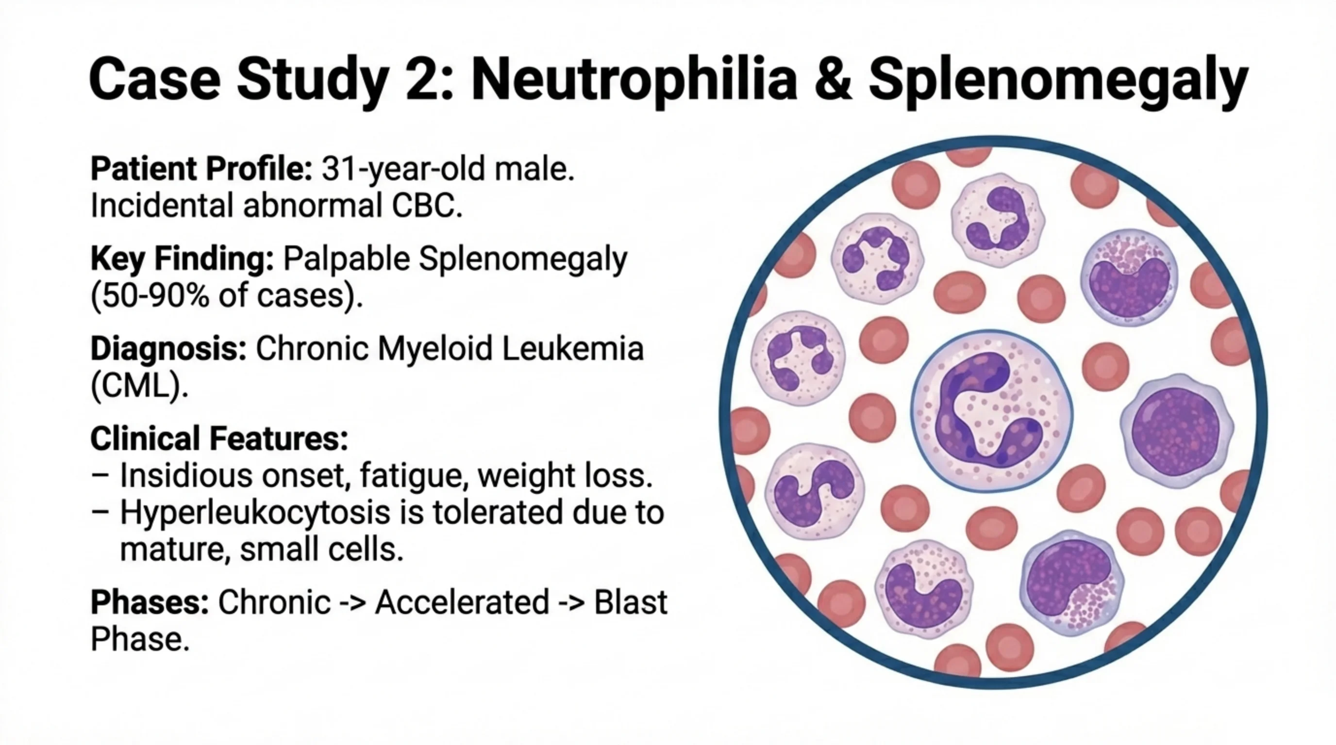
Case Study 2: Neutrophilia & Splenomegaly
Patient Profile: 31-year-old male. Incidental abnormal CBC.
Key Finding: Palpable Splenomegaly (50-90% of cases).
Diagnosis: Chronic Myeloid Leukemia (CML).
Clinical Features:
- Insidious onset, fatigue, weight loss.
- Hyperleukocytosis is tolerated due to mature, small cells.
Phases: Chronic -> Accelerated -> Blast Phase.
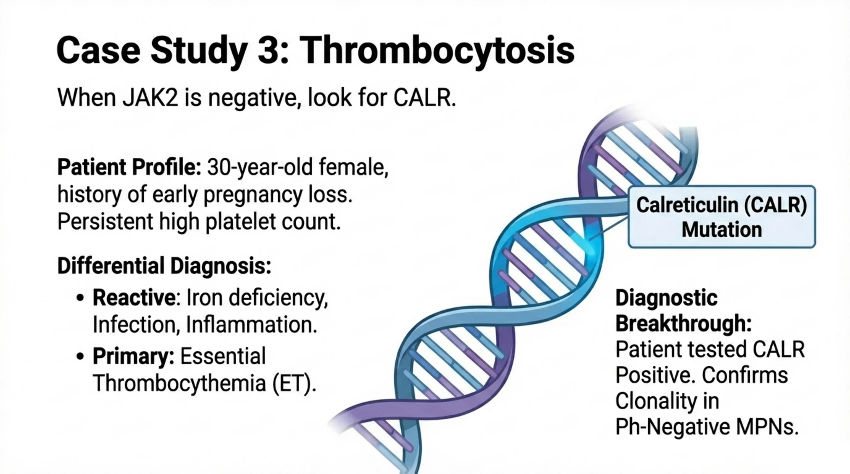
Case Study 3: Thrombocytosis
When JAK2 is negative, look for CALR.
Patient Profile: 30-year-old female, history of early pregnancy loss. Persistent high platelet count.
Differential Diagnosis:
- Reactive: Iron deficiency, Infection, Inflammation.
- Primary: Essential Thrombocythemia (ET).
Calreticulin (CALR) Mutation.
Diagnostic Breakthrough: Patient tested CALR Positive. Confirms Clonality in Ph-Negative MPNs.
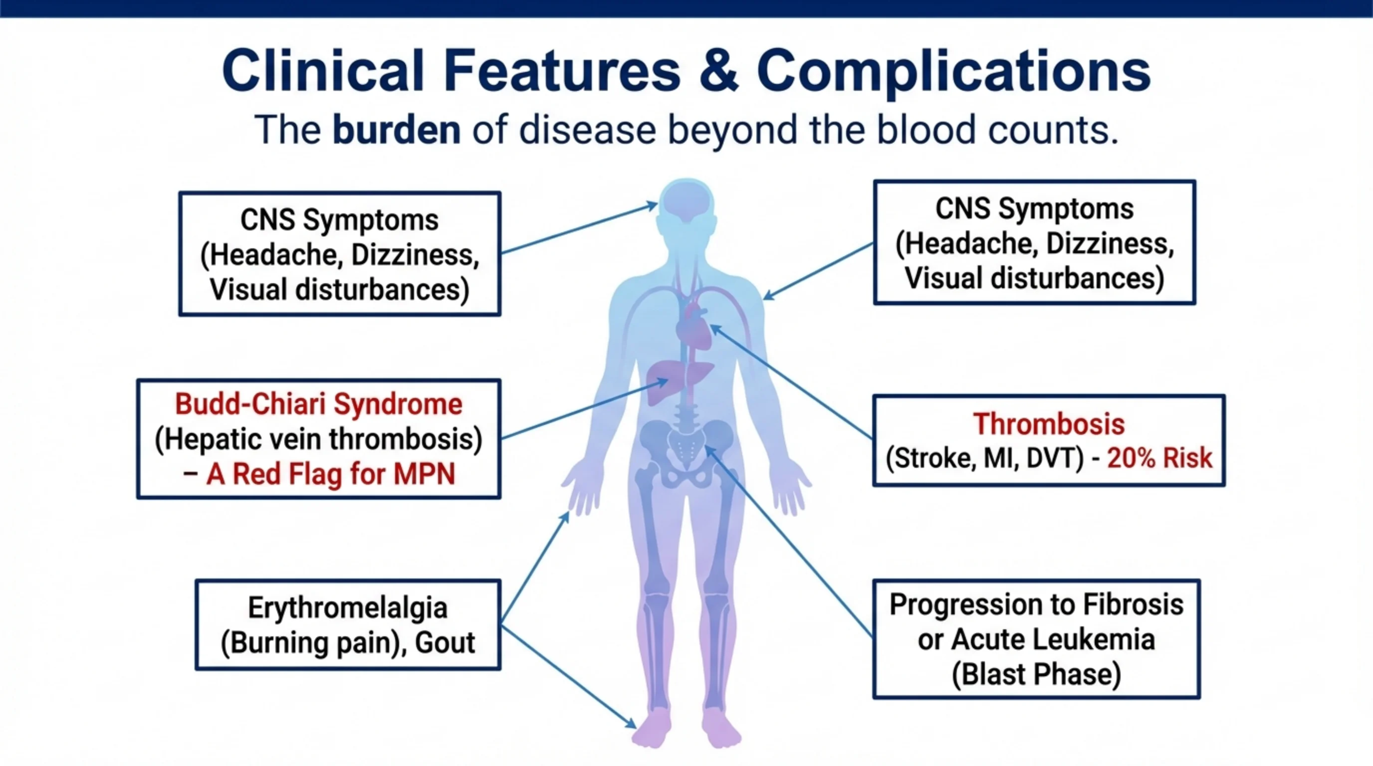
Clinical Features & Complications
The burden of disease beyond the blood counts.
- CNS Symptoms (Headache, Dizziness, Visual disturbances)
- Budd-Chiari Syndrome (Hepatic vein thrombosis) – A Red Flag for MPN
- Erythromelalgia (Burning pain), Gout
- Thrombosis (Stroke, MI, DVT) - 20% Risk
- Progression to Fibrosis or Acute Leukemia (Blast Phase)
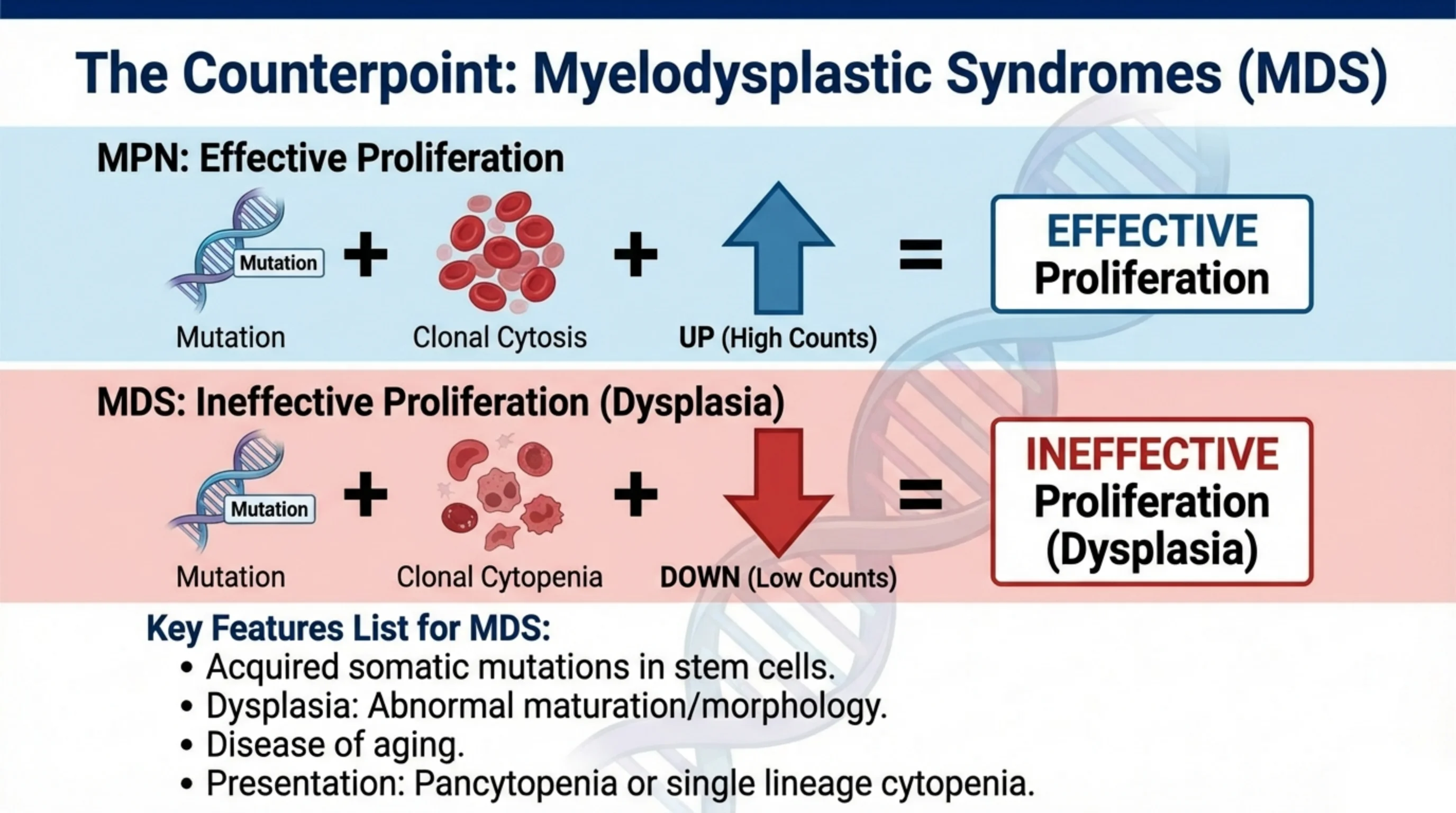
The Counterpoint: Myelodysplastic Syndromes (MDS)
MPN: Effective Proliferation — Mutation + Clonal Cytosis + UP (High Counts) = EFFECTIVE Proliferation.
MDS: Ineffective Proliferation (Dysplasia) — Mutation + Clonal Cytopenia + DOWN (Low Counts) = INEFFECTIVE Proliferation (Dysplasia).
Key Features List for MDS:
- Acquired somatic mutations in stem cells.
- Dysplasia: Abnormal maturation/morphology.
- Disease of aging.
- Presentation: Pancytopenia or single lineage cytopenia.
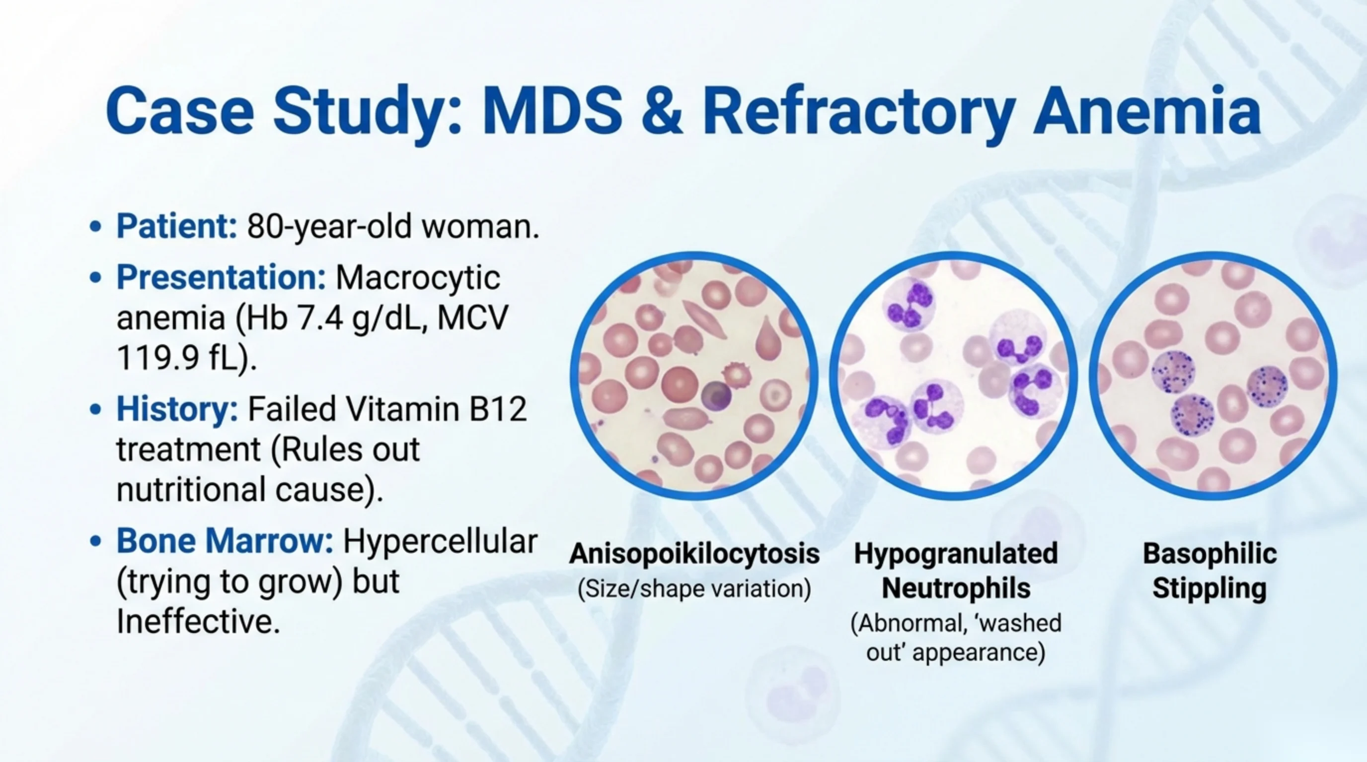
Case Study: MDS & Refractory Anemia
- Patient: 80-year-old woman.
- Presentation: Macrocytic anemia (Hb 7.4 g/dL, MCV 119.9 fL).
- History: Failed Vitamin B12 treatment (Rules out nutritional cause).
- Bone Marrow: Hypercellular (trying to grow) but Ineffective.
Blood smear findings:
- Anisopoikilocytosis (Size/shape variation)
- Hypogranulated Neutrophils (Abnormal, ‘washed out’ appearance)
- Basophilic Stippling
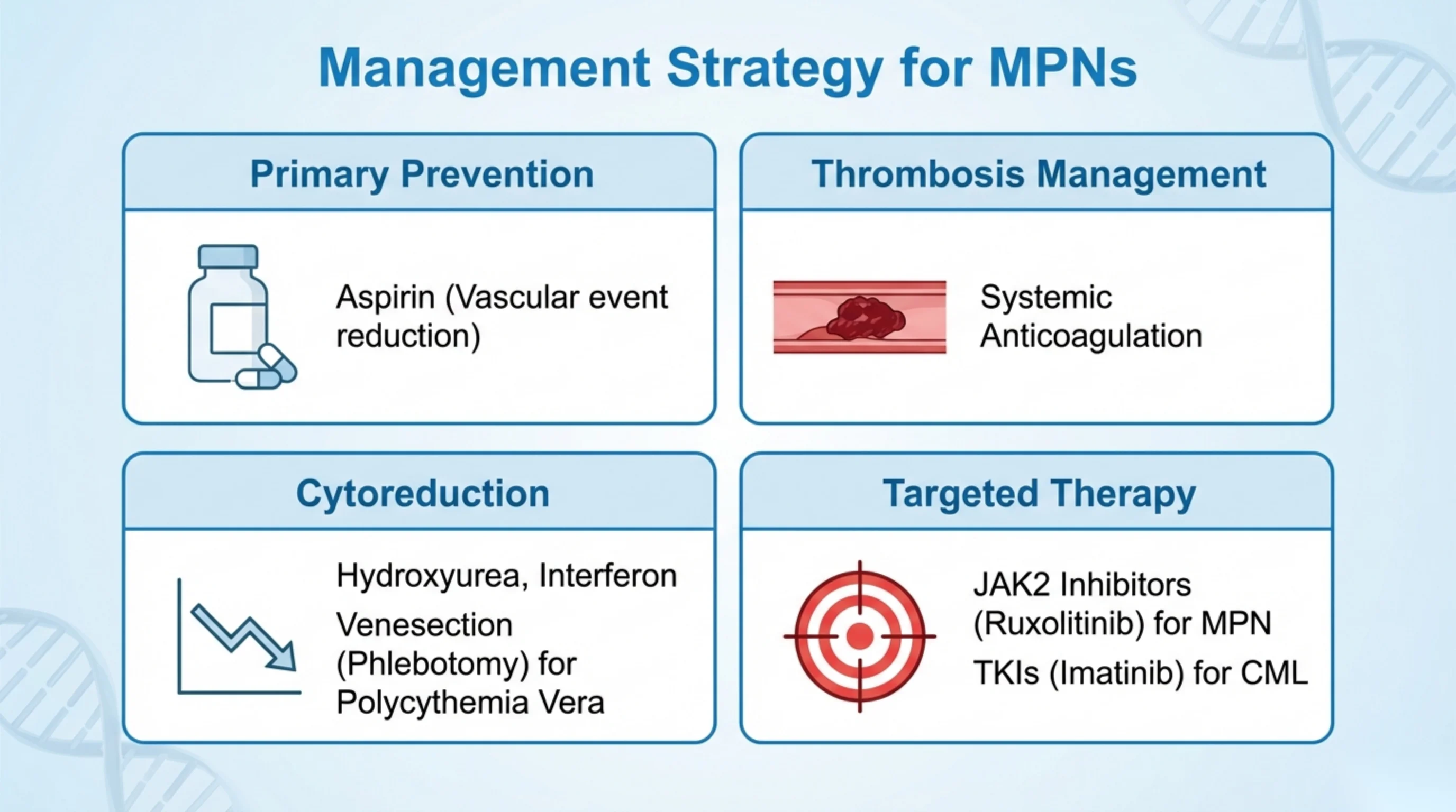
Management Strategy for MPNs
Primary Prevention
Aspirin (Vascular event reduction)
Thrombosis Management
Systemic Anticoagulation
Cytoreduction
Hydroxyurea, Interferon. Venesection (Phlebotomy) for Polycythemia Vera.
Targeted Therapy
JAK2 Inhibitors (Ruxolitinib) for MPN. TKIs (Imatinib) for CML.
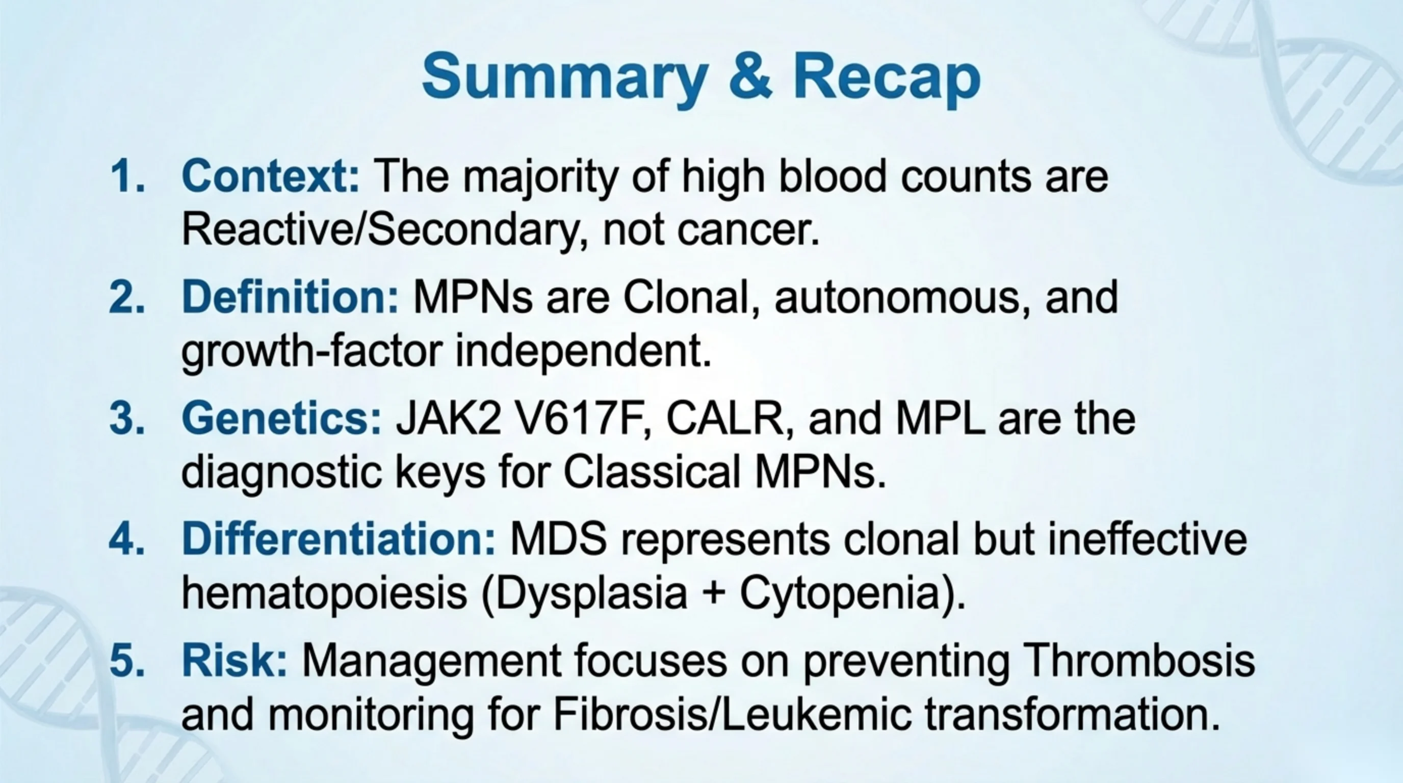
Summary & Recap
- Context: The majority of high blood counts are Reactive/Secondary, not cancer.
- Definition: MPNs are Clonal, autonomous, and growth-factor independent.
- Genetics: JAK2 V617F, CALR, and MPL are the diagnostic keys for Classical MPNs.
- Differentiation: MDS represents clonal but ineffective hematopoiesis (Dysplasia + Cytopenia).
- Risk: Management focuses on preventing Thrombosis and monitoring for Fibrosis/Leukemic transformation.

Questions & References
Contact: halkhaldy@kku.edu.sa
Thank you.